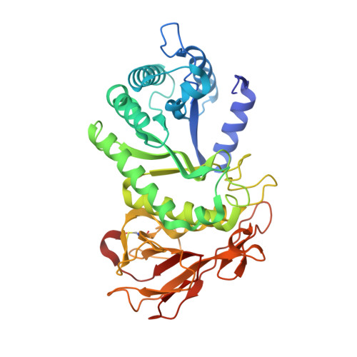
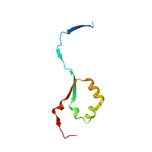
5E8M

Crystal structure of human heparanase


Experimental Data Snapshot

  • Method: X-RAY DIFFRACTION
  • Resolution: 1.75 Å
  • R-Value Free: 0.200 
  • R-Value Work: 0.164 
  • R-Value Observed: 0.166 

wwPDB Validation   3D Report Full Report


Ligand Structure Quality Assessment 


This is version 2.1 of the entry. See complete history


Literature

Structural characterization of human heparanase reveals insights into substrate recognition.

Wu, L.Viola, C.M.Brzozowski, A.M.Davies, G.J.

(2015) Nat Struct Mol Biol 22: 1016-1022

  • DOI: https://doi.org/10.1038/nsmb.3136
  • Primary Citation of Related Structures:  
    5E8M, 5E97, 5E98, 5E9B, 5E9C

  • PubMed Abstract: 

    Heparan sulfate (HS) is a glycosaminoglycan that forms a key component of the extracellular matrix (ECM). Breakdown of HS is carried out by heparanase (HPSE), an endo-β-glucuronidase of the glycoside hydrolase 79 (GH79) family. Overexpression of HPSE results in breakdown of extracellular HS and release of stored growth factors and hence is strongly linked to cancer metastasis. Here we present crystal structures of human HPSE at 1.6-Å to 1.9-Å resolution that reveal how an endo-acting binding cleft is exposed by proteolytic activation of latent proHPSE. We used oligosaccharide complexes to map the substrate-binding and sulfate-recognition motifs. These data shed light on the structure and interactions of a key enzyme involved in ECM maintenance and provide a starting point for the design of HPSE inhibitors for use as biochemical tools and anticancer therapeutics.


  • Organizational Affiliation

    Department of Chemistry, University of York, York, UK.


Macromolecules
Find similar proteins by:  (by identity cutoff)  |  3D Structure
Entity ID: 1
MoleculeChains Sequence LengthOrganismDetailsImage
Heparanase389Homo sapiensMutation(s): 1 
Gene Names: HPSEHEPHPAHPA1HPR1HPSE1HSE1
EC: 3.2.1.166
UniProt & NIH Common Fund Data Resources
Find proteins for Q9Y251 (Homo sapiens)
Explore Q9Y251 
Go to UniProtKB:  Q9Y251
PHAROS:  Q9Y251
GTEx:  ENSG00000173083 
Entity Groups  
Sequence Clusters30% Identity50% Identity70% Identity90% Identity95% Identity100% Identity
UniProt GroupQ9Y251
Glycosylation
Glycosylation Sites: 5Go to GlyGen: Q9Y251-1
Sequence Annotations
Expand
  • Reference Sequence
Find similar proteins by:  (by identity cutoff)  |  3D Structure
Entity ID: 2
MoleculeChains Sequence LengthOrganismDetailsImage
Heparanase77Homo sapiensMutation(s): 0 
Gene Names: HPSEHEPHPAHPA1HPR1HPSE1HSE1
EC: 3.2.1.166
UniProt & NIH Common Fund Data Resources
Find proteins for Q9Y251 (Homo sapiens)
Explore Q9Y251 
Go to UniProtKB:  Q9Y251
PHAROS:  Q9Y251
GTEx:  ENSG00000173083 
Entity Groups  
Sequence Clusters30% Identity50% Identity70% Identity90% Identity95% Identity100% Identity
UniProt GroupQ9Y251
Sequence Annotations
Expand
  • Reference Sequence
Oligosaccharides

Help

Entity ID: 3
MoleculeChains Length2D Diagram Glycosylation3D Interactions
alpha-L-fucopyranose-(1-6)-2-acetamido-2-deoxy-beta-D-glucopyranose
C
2N-Glycosylation
Glycosylation Resources
GlyTouCan:  G86851RC
GlyCosmos:  G86851RC
GlyGen:  G86851RC
Small Molecules
Ligands 3 Unique
IDChains Name / Formula / InChI Key2D Diagram3D Interactions
NAG
Query on NAG

Download Ideal Coordinates CCD File 
D [auth A],
E [auth A],
F [auth A],
G [auth A]
2-acetamido-2-deoxy-beta-D-glucopyranose
C8 H15 N O6
OVRNDRQMDRJTHS-FMDGEEDCSA-N
EDO
Query on EDO

Download Ideal Coordinates CCD File 
H [auth A]
I [auth A]
J [auth A]
K [auth A]
L [auth A]
H [auth A],
I [auth A],
J [auth A],
K [auth A],
L [auth A],
M [auth A],
N [auth A],
O [auth A],
P [auth A],
Q [auth A],
R [auth A],
S [auth A],
T [auth A],
U [auth A],
Y [auth B]
1,2-ETHANEDIOL
C2 H6 O2
LYCAIKOWRPUZTN-UHFFFAOYSA-N
CL
Query on CL

Download Ideal Coordinates CCD File 
V [auth A],
W [auth A],
X [auth A]
CHLORIDE ION
Cl
VEXZGXHMUGYJMC-UHFFFAOYSA-M
Experimental Data & Validation

Experimental Data

  • Method: X-RAY DIFFRACTION
  • Resolution: 1.75 Å
  • R-Value Free: 0.200 
  • R-Value Work: 0.164 
  • R-Value Observed: 0.166 
  • Space Group: P 1 21 1
Unit Cell:
Length ( Å )Angle ( ˚ )
a = 46.765α = 90
b = 70.972β = 94.17
c = 78.948γ = 90
Software Package:
Software NamePurpose
REFMACrefinement
XDSdata reduction
Aimlessdata scaling
SHELXphasing

Structure Validation

View Full Validation Report


Ligand Structure Quality Assessment 


Entry History & Funding Information

Deposition Data


Funding OrganizationLocationGrant Number
European Research CouncilUnited Kingdom--

Revision History  (Full details and data files)

  • Version 1.0: 2015-11-18
    Type: Initial release
  • Version 1.1: 2015-12-02
    Changes: Database references
  • Version 1.2: 2015-12-16
    Changes: Database references
  • Version 1.3: 2016-05-04
    Changes: Structure summary
  • Version 2.0: 2020-07-29
    Type: Remediation
    Reason: Carbohydrate remediation
    Changes: Atomic model, Data collection, Derived calculations, Structure summary
  • Version 2.1: 2024-11-20
    Changes: Data collection, Database references, Derived calculations, Structure summary