5EMY | pdb_00005emy

Human Pancreatic Alpha-Amylase in complex with the mechanism based inactivator glucosyl epi-cyclophellitol


Experimental Data Snapshot

  • Method: X-RAY DIFFRACTION
  • Resolution: 1.23 Å
  • R-Value Free: 
    0.142 (Depositor), 0.144 (DCC) 
  • R-Value Work: 
    0.119 (Depositor), 0.120 (DCC) 
  • R-Value Observed: 
    0.120 (Depositor) 

Starting Model: experimental
View more details

wwPDB Validation 3D Report Full Report

Validation slider image for 5EMY

Ligand Structure Quality Assessment 


This is version 3.2 of the entry. See complete history

Literature

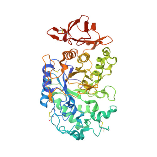
Glucosyl epi-cyclophellitol allows mechanism-based inactivation and structural analysis of human pancreatic alpha-amylase.

Caner, S.Zhang, X.Jiang, J.Chen, H.M.Nguyen, N.T.Overkleeft, H.Brayer, G.D.Withers, S.G.

(2016) FEBS Lett 590: 1143-1151

  • DOI: https://doi.org/10.1002/1873-3468.12143
  • Primary Citation Related Structures: 
    5EMY

  • PubMed Abstract: 

    As part of a search for selective, mechanism-based covalent inhibitors of human pancreatic α-amylase we describe the chemoenzymatic synthesis of the disaccharide analog α-glucosyl epi-cyclophellitol, demonstrate its stoichiometric reaction with human pancreatic α-amylase and evaluate the time dependence of its inhibition. X-ray crystallographic analysis of the covalent derivative so formed confirms its reaction at the active site with formation of a covalent bond to the catalytic nucleophile D197. The structure illuminates the interactions with the active site and confirms OH4' on the nonreducing end sugar as a good site for attachment of fluorescent tags in generating probes for localization and quantitation of amylase in vivo.


  • Organizational Affiliation
    • Department of Biochemistry and Molecular Biology, Faculty of Medicine, University of British Columbia, Vancouver, BC, Canada.

Macromolecule Content 

  • Total Structure Weight: 56.35 kDa 
  • Atom Count: 4,561 
  • Modeled Residue Count: 496 
  • Deposited Residue Count: 496 
  • Unique protein chains: 1

Macromolecules

Find similar proteins by:|  3D Structure
Entity ID: 1
MoleculeChains  Sequence LengthOrganismDetailsImage
Pancreatic alpha-amylase496Homo sapiensMutation(s): 0 
Gene Names: AMY2A
EC: 3.2.1.1
UniProt & NIH Common Fund Data Resources
Find proteins for P04746 (Homo sapiens)
Explore P04746 
Go to UniProtKB:  P04746
PHAROS:  P04746
GTEx:  ENSG00000243480 
Entity Groups
Sequence Clusters30% Identity50% Identity70% Identity90% Identity95% Identity100% Identity
UniProt GroupP04746
Sequence Annotations
Expand
Reference Sequence

Small Molecules

Modified Residues  1 Unique
IDChains TypeFormula2D DiagramParent
PCA
Query on PCA
A
L-PEPTIDE LINKINGC5 H7 N O3GLN

Experimental Data & Validation

Experimental Data

  • Method: X-RAY DIFFRACTION
  • Resolution: 1.23 Å
  • R-Value Free:  0.142 (Depositor), 0.144 (DCC) 
  • R-Value Work:  0.119 (Depositor), 0.120 (DCC) 
  • R-Value Observed: 0.120 (Depositor) 
Space Group: P 21 21 21
Unit Cell:
Length ( Å )Angle ( ˚ )
a = 52.33α = 90
b = 68.18β = 90
c = 130.38γ = 90
Software Package:
Software NamePurpose
PHENIXrefinement
XDSdata reduction
XSCALEdata scaling
PHASERphasing

Structure Validation

View Full Validation Report


Ligand Structure Quality Assessment 


Entry History 

& Funding Information

Deposition Data


Funding OrganizationLocationGrant Number
Canadian Institutes of Health Research (CIHR)CanadaCIHR MOP-111082

Revision History  (Full details and data files)

  • Version 1.0: 2016-07-06
    Type: Initial release
  • Version 2.0: 2020-01-08
    Changes: Author supporting evidence, Database references, Derived calculations, Polymer sequence
  • Version 3.0: 2020-07-29
    Type: Remediation
    Reason: Carbohydrate remediation
    Changes: Atomic model, Derived calculations, Structure summary
  • Version 3.1: 2023-09-27
    Changes: Data collection, Database references, Refinement description, Structure summary
  • Version 3.2: 2024-10-23
    Changes: Structure summary