5EWK

Scabin toxin from Streptomyces Scabies in complex with inhibitor PJ34


Experimental Data Snapshot

  • Method: X-RAY DIFFRACTION
  • Resolution: 1.60 Å
  • R-Value Free: 
    0.179 (Depositor), 0.180 (DCC) 
  • R-Value Work: 
    0.154 (Depositor), 0.150 (DCC) 
  • R-Value Observed: 
    0.155 (Depositor) 

Starting Model: experimental
View more details

wwPDB Validation   3D Report Full Report


Ligand Structure Quality Assessment 

Created with Raphaël 2.3.0Worse 01 BetterLigand structure goodness of fit to experimental dataBest fitted P34Click on this verticalbar to view details

This is version 1.5 of the entry. See complete history


Literature

Scabin, a Novel DNA-acting ADP-ribosyltransferase from Streptomyces scabies.

Lyons, B.Ravulapalli, R.Lanoue, J.Lugo, M.R.Dutta, D.Carlin, S.Merrill, A.R.

(2016) J Biol Chem 291: 11198-11215

  • DOI: https://doi.org/10.1074/jbc.M115.707653
  • Primary Citation of Related Structures:  
    5DAZ, 5EWK, 5EWY

  • PubMed Abstract: 

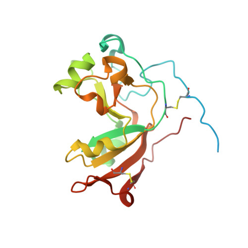
    A bioinformatics strategy was used to identify Scabin, a novel DNA-targeting enzyme from the plant pathogen 87.22 strain of Streptomyces scabies Scabin shares nearly 40% sequence identity with the Pierisin family of mono-ADP-ribosyltransferase toxins. Scabin was purified to homogeneity as a 22-kDa single-domain enzyme and was shown to possess high NAD(+)-glycohydrolase (Km (NAD) = 68 ± 3 μm; kcat = 94 ± 2 min(-1)) activity with an RSQXE motif; it was also shown to target deoxyguanosine and showed sigmoidal enzyme kinetics (K0.5(deoxyguanosine) = 302 ± 12 μm; kcat = 14 min(-1)). Mass spectrometry analysis revealed that Scabin labels the exocyclic amino group on guanine bases in either single-stranded or double-stranded DNA. Several small molecule inhibitors were identified, and the most potent compounds were found to inhibit the enzyme activity with Ki values ranging from 3 to 24 μm PJ34, a well known inhibitor of poly-ADP-ribosyltransferases, was shown to be the most potent inhibitor of Scabin. Scabin was crystallized, representing the first structure of a DNA-targeting mono-ADP-ribosyltransferase enzyme; the structures of the apo-form (1.45 Å) and with two inhibitors (P6-E, 1.4 Å; PJ34, 1.6 Å) were solved. These x-ray structures are also the first high resolution structures of the Pierisin subgroup of the mono-ADP-ribosyltransferase toxin family. A model of Scabin with its DNA substrate is also proposed.


  • Organizational Affiliation

    From the Department of Molecular and Cellular Biology, University of Guelph, Guelph, Ontario N1G 2W1, Canada and.


Macromolecules
Find similar proteins by:  (by identity cutoff)  |  3D Structure
Entity ID: 1
MoleculeChains Sequence LengthOrganismDetailsImage
Putative ADP-Ribosyltransferase Scabin200Streptomyces scabiei 87.22Mutation(s): 0 
Gene Names: SCAB_27771
EC: 2.4.2.31
UniProt
Find proteins for C9Z6T8 (Streptomyces scabiei (strain 87.22))
Explore C9Z6T8 
Go to UniProtKB:  C9Z6T8
Entity Groups  
Sequence Clusters30% Identity50% Identity70% Identity90% Identity95% Identity100% Identity
UniProt GroupC9Z6T8
Sequence Annotations
Expand
  • Reference Sequence
Small Molecules
Ligands 1 Unique
IDChains Name / Formula / InChI Key2D Diagram3D Interactions
P34
Query on P34

Download Ideal Coordinates CCD File 
B [auth A]N~2~,N~2~-DIMETHYL-N~1~-(6-OXO-5,6-DIHYDROPHENANTHRIDIN-2-YL)GLYCINAMIDE
C17 H17 N3 O2
UYJZZVDLGDDTCL-UHFFFAOYSA-N
Experimental Data & Validation

Experimental Data

  • Method: X-RAY DIFFRACTION
  • Resolution: 1.60 Å
  • R-Value Free:  0.179 (Depositor), 0.180 (DCC) 
  • R-Value Work:  0.154 (Depositor), 0.150 (DCC) 
  • R-Value Observed: 0.155 (Depositor) 
Space Group: C 1 2 1
Unit Cell:
Length ( Å )Angle ( ˚ )
a = 87.47α = 90
b = 61.18β = 100.38
c = 38.03γ = 90
Software Package:
Software NamePurpose
PHENIXrefinement
XDSdata scaling

Structure Validation

View Full Validation Report


Ligand Structure Quality Assessment 

Created with Raphaël 2.3.0Worse 01 BetterLigand structure goodness of fit to experimental dataBest fitted P34Click on this verticalbar to view details

Entry History 

Deposition Data

Revision History  (Full details and data files)

  • Version 1.0: 2016-03-23
    Type: Initial release
  • Version 1.1: 2016-04-06
    Changes: Database references
  • Version 1.2: 2016-06-08
    Changes: Database references
  • Version 1.3: 2017-11-22
    Changes: Advisory, Database references, Derived calculations, Refinement description
  • Version 1.4: 2023-09-27
    Changes: Advisory, Data collection, Database references, Refinement description
  • Version 1.5: 2024-10-23
    Changes: Structure summary