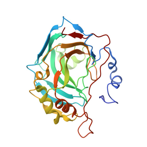
5G01

An unusual natural product primary sulfonamide: synthesis, carbonic anhydrase inhibition and protein x-ray structure of Psammaplin C


Experimental Data Snapshot

  • Method: X-RAY DIFFRACTION
  • Resolution: 1.40 Å
  • R-Value Free: 0.165 
  • R-Value Work: 0.128 
  • R-Value Observed: 0.130 

Starting Model: experimental
View more details

wwPDB Validation   3D Report Full Report


Ligand Structure Quality Assessment 


This is version 1.3 of the entry. See complete history


Literature

An Unusual Natural Product Primary Sulfonamide: Synthesis, Carbonic Anhydrase Inhibition and Protein X-Ray Structures of Psammaplin C.

Mujumdar, P.Teruya, K.Tonissen, K.F.Vullo, D.Supuran, C.T.Peat, T.S.Poulsen, S.

(2016) J Med Chem 59: 5462

  • DOI: https://doi.org/10.1021/acs.jmedchem.6b00443
  • Primary Citation of Related Structures:  
    5A6H, 5G01, 5G03, 5G0B, 5G0C

  • PubMed Abstract: 

    Psammaplin C is one of only two described natural product primary sulfonamides. Here we report the synthesis of psammaplin C and evaluate the inhibition profile against therapeutically relevant carbonic anhydrase (CA) zinc metalloenzymes. The compound exhibited unprecedented inhibition of an important cancer-associated isozyme, hCA XII, with a Ki of 0.79 nM. The compound also displayed good isoform selectivity for hCA XII over other CAs. We present the first reported protein X-ray crystal structures of psammaplin C in complex with human CAs. We engineered the easily crystallized hCA II enzyme to mimic both the hCA IX and hCA XII binding sites and then utilized protein X-ray crystallography to determine the binding pose of psammaplin C within the hCA II, hCA IX, and hCA XII mimic active sites, all to high resolution. This is the first time a natural product primary sulfonamide inhibitor has been assessed for inhibition and binding to CAs.


  • Organizational Affiliation

    Eskitis Institute for Drug Discovery, Griffith University , Don Young Road, Nathan, Queensland 4111, Australia.


Macromolecules
Find similar proteins by:  (by identity cutoff)  |  3D Structure
Entity ID: 1
MoleculeChains Sequence LengthOrganismDetailsImage
CARBONIC ANHYDRASE 2260Homo sapiensMutation(s): 9 
EC: 4.2.1.1 (PDB Primary Data), 4.2.1.69 (UniProt)
UniProt & NIH Common Fund Data Resources
Find proteins for P00918 (Homo sapiens)
Explore P00918 
Go to UniProtKB:  P00918
PHAROS:  P00918
GTEx:  ENSG00000104267 
Entity Groups  
Sequence Clusters30% Identity50% Identity70% Identity90% Identity95% Identity100% Identity
UniProt GroupP00918
Sequence Annotations
Expand
  • Reference Sequence
Experimental Data & Validation

Experimental Data

  • Method: X-RAY DIFFRACTION
  • Resolution: 1.40 Å
  • R-Value Free: 0.165 
  • R-Value Work: 0.128 
  • R-Value Observed: 0.130 
  • Space Group: P 1 21 1
Unit Cell:
Length ( Å )Angle ( ˚ )
a = 42.159α = 90
b = 41.494β = 104.26
c = 71.699γ = 90
Software Package:
Software NamePurpose
REFMACrefinement
XDSdata reduction
Aimlessdata scaling
PHASERphasing

Structure Validation

View Full Validation Report


Ligand Structure Quality Assessment 


Entry History 

Deposition Data

Revision History  (Full details and data files)

  • Version 1.0: 2016-05-25
    Type: Initial release
  • Version 1.1: 2016-06-22
    Changes: Database references
  • Version 1.2: 2019-05-08
    Changes: Data collection, Experimental preparation, Other
  • Version 1.3: 2024-01-10
    Changes: Data collection, Database references, Derived calculations, Other, Refinement description