5HR6

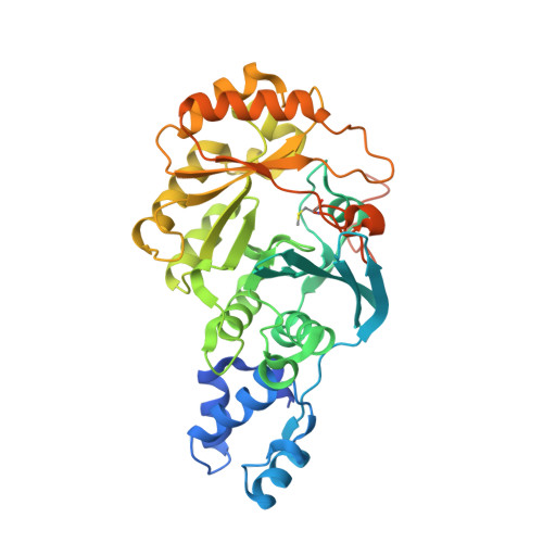
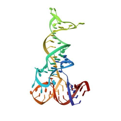
X-ray crystal structure of C118A RlmN with cross-linked tRNA purified from Escherichia coli


Experimental Data Snapshot

  • Method: X-RAY DIFFRACTION
  • Resolution: 2.88 Å
  • R-Value Free: 0.228 
  • R-Value Work: 0.182 
  • R-Value Observed: 0.184 

wwPDB Validation   3D Report Full Report


Ligand Structure Quality Assessment 


This is version 1.2 of the entry. See complete history


Literature

Crystallographic capture of a radical S-adenosylmethionine enzyme in the act of modifying tRNA.

Schwalm, E.L.Grove, T.L.Booker, S.J.Boal, A.K.

(2016) Science 352: 309-312

  • DOI: https://doi.org/10.1126/science.aad5367
  • Primary Citation of Related Structures:  
    5HR6, 5HR7

  • PubMed Abstract: 

    RlmN is a dual-specificity RNA methylase that modifies C2 of adenosine 2503 (A2503) in 23S rRNA and C2 of adenosine 37 (A37) in several Escherichia coli transfer RNAs (tRNAs). A related methylase, Cfr, modifies C8 of A2503 via a similar mechanism, conferring resistance to multiple classes of antibiotics. Here, we report the x-ray structure of a key intermediate in the RlmN reaction, in which a Cys(118)→Ala variant of the protein is cross-linked to a tRNA(Glu)substrate through the terminal methylene carbon of a formerly methylcysteinyl residue and C2 of A37. RlmN contacts the entire length of tRNA(Glu), accessing A37 by using an induced-fit strategy that completely unfolds the tRNA anticodon stem-loop, which is likely critical for recognition of both tRNA and ribosomal RNA substrates.


  • Organizational Affiliation

    Department of Chemistry, Pennsylvania State University, University Park, PA 16802, USA.


Macromolecules

Find similar proteins by:  (by identity cutoff)  |  3D Structure
Entity ID: 1
MoleculeChains Sequence LengthOrganismDetailsImage
RlmN methylaseA,
C [auth B]
404Escherichia coliMutation(s): 1 
EC: 2.1.1.192
UniProt
Find proteins for P36979 (Escherichia coli (strain K12))
Explore P36979 
Go to UniProtKB:  P36979
Entity Groups  
Sequence Clusters30% Identity50% Identity70% Identity90% Identity95% Identity100% Identity
UniProt GroupP36979
Sequence Annotations
Expand
  • Reference Sequence
Find similar nucleic acids by:  (by identity cutoff)  |  3D Structure
Entity ID: 2
MoleculeChains LengthOrganismImage
tRNA GluB [auth C],
D
68Escherichia coli
Sequence Annotations
Expand
  • Reference Sequence
Small Molecules
Ligands 4 Unique
IDChains Name / Formula / InChI Key2D Diagram3D Interactions
SF4
Query on SF4

Download Ideal Coordinates CCD File 
F [auth A],
M [auth B]
IRON/SULFUR CLUSTER
Fe4 S4
LJBDFODJNLIPKO-UHFFFAOYSA-N
5AD
Query on 5AD

Download Ideal Coordinates CCD File 
G [auth A],
N [auth B]
5'-DEOXYADENOSINE
C10 H13 N5 O3
XGYIMTFOTBMPFP-KQYNXXCUSA-N
MET
Query on MET

Download Ideal Coordinates CCD File 
E [auth A],
L [auth B]
METHIONINE
C5 H11 N O2 S
FFEARJCKVFRZRR-BYPYZUCNSA-N
MG
Query on MG

Download Ideal Coordinates CCD File 
H [auth A]
I [auth C]
J [auth C]
K [auth C]
O [auth B]
H [auth A],
I [auth C],
J [auth C],
K [auth C],
O [auth B],
P [auth D]
MAGNESIUM ION
Mg
JLVVSXFLKOJNIY-UHFFFAOYSA-N
Modified Residues  1 Unique
IDChains TypeFormula2D DiagramParent
SMC
Query on SMC
A,
C [auth B]
L-PEPTIDE LINKINGC4 H9 N O2 SCYS
Experimental Data & Validation

Experimental Data

  • Method: X-RAY DIFFRACTION
  • Resolution: 2.88 Å
  • R-Value Free: 0.228 
  • R-Value Work: 0.182 
  • R-Value Observed: 0.184 
  • Space Group: P 1 21 1
Unit Cell:
Length ( Å )Angle ( ˚ )
a = 88.664α = 90
b = 69.821β = 90.3
c = 149.298γ = 90
Software Package:
Software NamePurpose
REFMACrefinement
HKL-2000data scaling
PDB_EXTRACTdata extraction
HKL-2000data reduction
PHASERphasing

Structure Validation

View Full Validation Report


Ligand Structure Quality Assessment 


Entry History 

Deposition Data

Revision History  (Full details and data files)

  • Version 1.0: 2016-04-13
    Type: Initial release
  • Version 1.1: 2016-04-27
    Changes: Database references
  • Version 1.2: 2024-11-13
    Changes: Data collection, Database references, Derived calculations, Structure summary