Nucleotide binding by the widespread high-affinity cyclic di-GMP receptor MshEN domain.
Wang, Y.C., Chin, K.H., Tu, Z.L., He, J., Jones, C.J., Sanchez, D.Z., Yildiz, F.H., Galperin, M.Y., Chou, S.H.(2016) Nat Commun 7: 12481-12481
- PubMed: 27578558 Search on PubMedSearch on PubMed Central
- DOI: https://doi.org/10.1038/ncomms12481
- Primary Citation Related Structures:
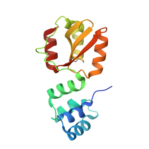
5HTL - PubMed Abstract:
C-di-GMP is a bacterial second messenger regulating various cellular functions. Many bacteria contain c-di-GMP-metabolizing enzymes but lack known c-di-GMP receptors. Recently, two MshE-type ATPases associated with bacterial type II secretion system and type IV pilus formation were shown to specifically bind c-di-GMP. Here we report crystal structure of the MshE N-terminal domain (MshEN1-145) from Vibrio cholerae in complex with c-di-GMP at a 1.37 Å resolution. This structure reveals a unique c-di-GMP-binding mode, featuring a tandem array of two highly conserved binding motifs, each comprising a 24-residue sequence RLGxx(L/V/I)(L/V/I)xxG(L/V/I)(L/V/I)xxxxLxxxLxxQ that binds half of the c-di-GMP molecule, primarily through hydrophobic interactions. Mutating these highly conserved residues markedly reduces c-di-GMP binding and biofilm formation by V. cholerae. This c-di-GMP-binding motif is present in diverse bacterial proteins exhibiting binding affinities ranging from 0.5 μM to as low as 14 nM. The MshEN domain contains the longest nucleotide-binding motif reported to date.
- Institute of Biochemistry, National Chung Hsing University, Taichung 40227, Taiwan, Republic of China.
Organizational Affiliation: