5I42

Structure of HIV-1 Reverse Transcriptase in complex with a DNA aptamer, AZTTP, and CA(2+) ion


Experimental Data Snapshot

  • Method: X-RAY DIFFRACTION
  • Resolution: 3.30 Å
  • R-Value Free: 
    0.247 (Depositor), 0.250 (DCC) 
  • R-Value Work: 
    0.221 (Depositor), 0.220 (DCC) 
  • R-Value Observed: 
    0.222 (Depositor) 

Starting Model: experimental
View more details

wwPDB Validation   3D Report Full Report


Ligand Structure Quality Assessment 

Created with Raphaël 2.3.0Worse 01 BetterLigand structure goodness of fit to experimental dataBest fitted AZTClick on this verticalbar to view details

This is version 1.5 of the entry. See complete history


Literature

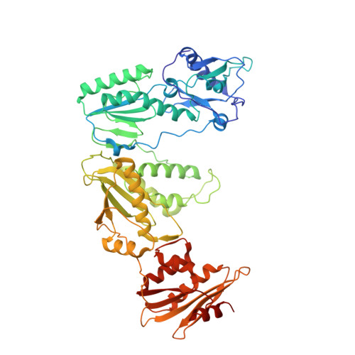
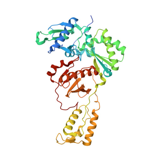
Conformational States of HIV-1 Reverse Transcriptase for Nucleotide Incorporation vs Pyrophosphorolysis-Binding of Foscarnet.

Das, K.Balzarini, J.Miller, M.T.Maguire, A.R.DeStefano, J.J.Arnold, E.

(2016) ACS Chem Biol 11: 2158-2164

  • DOI: https://doi.org/10.1021/acschembio.6b00187
  • Primary Citation of Related Structures:  
    5HP1, 5HRO, 5I3U, 5I42

  • PubMed Abstract: 

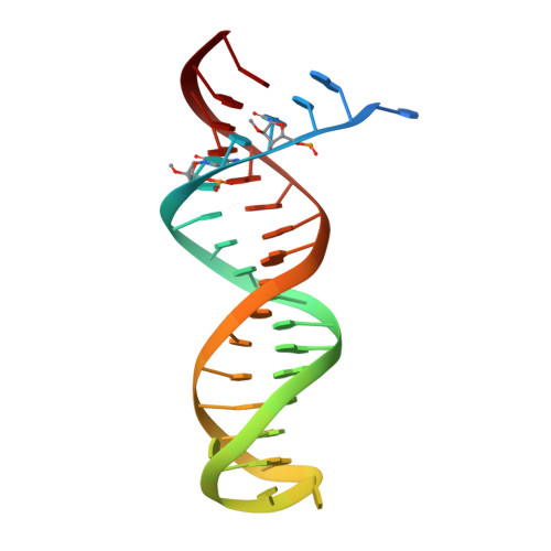
    HIV-1 reverse transcriptase (RT) catalytically incorporates individual nucleotides into a viral DNA strand complementing an RNA or DNA template strand; the polymerase active site of RT adopts multiple conformational and structural states while performing this task. The states associated are dNTP binding at the N site, catalytic incorporation of a nucleotide, release of a pyrophosphate, and translocation of the primer 3'-end to the P site. Structural characterization of each of these states may help in understanding the molecular mechanisms of drug activity and resistance and in developing new RT inhibitors. Using a 38-mer DNA template-primer aptamer as the substrate mimic, we crystallized an RT/dsDNA complex that is catalytically active, yet translocation-incompetent in crystals. The ability of RT to perform dNTP binding and incorporation in crystals permitted obtaining a series of structures: (I) RT/DNA (P-site), (II) RT/DNA/AZTTP ternary, (III) RT/AZT-terminated DNA (N-site), and (IV) RT/AZT-terminated DNA (N-site)/foscarnet complexes. The stable N-site complex permitted the binding of foscarnet as a pyrophosphate mimic. The Mg(2+) ions dissociated after catalytic addition of AZTMP in the pretranslocated structure III, whereas ions A and B had re-entered the active site to bind foscarnet in structure IV. The binding of foscarnet involves chelation with the Mg(2+) (B) ion and interactions with K65 and R72. The analysis of interactions of foscarnet and the recently discovered nucleotide-competing RT inhibitor (NcRTI) α-T-CNP in two different conformational states of the enzyme provides insights for developing new classes of polymerase active site RT inhibitors.


  • Organizational Affiliation

    Center for Advanced Biotechnology and Medicine (CABM), Department of Chemistry and Chemical Biology, Rutgers University , Piscataway, New Jersey, United States.


Macromolecules

Find similar proteins by:  (by identity cutoff)  |  3D Structure
Entity ID: 1
MoleculeChains Sequence LengthOrganismDetailsImage
HIV-1 REVERSE TRANSCRIPTASE P66 SUBUNIT
A, C
555Human immunodeficiency virus type 1 BH10Mutation(s): 1 
Gene Names: gag-pol
EC: 2.7.7.49
UniProt
Find proteins for P03366 (Human immunodeficiency virus type 1 group M subtype B (isolate BH10))
Explore P03366 
Go to UniProtKB:  P03366
Entity Groups  
Sequence Clusters30% Identity50% Identity70% Identity90% Identity95% Identity100% Identity
UniProt GroupP03366
Sequence Annotations
Expand
  • Reference Sequence
Find similar proteins by:  (by identity cutoff)  |  3D Structure
Entity ID: 2
MoleculeChains Sequence LengthOrganismDetailsImage
HIV-1 REVERSE TRANSCRIPTASE P51 SUBUNIT
B, D
444Human immunodeficiency virus type 1 BH10Mutation(s): 1 
Gene Names: gag-pol
EC: 2.7.7.49
UniProt
Find proteins for P03366 (Human immunodeficiency virus type 1 group M subtype B (isolate BH10))
Explore P03366 
Go to UniProtKB:  P03366
Entity Groups  
Sequence Clusters30% Identity50% Identity70% Identity90% Identity95% Identity100% Identity
UniProt GroupP03366
Sequence Annotations
Expand
  • Reference Sequence
Small Molecules
Binding Affinity Annotations 
IDSourceBinding Affinity
AZT BindingDB:  5I42 Ki: min: 47, max: 91 (nM) from 2 assay(s)
Kd: min: 1150, max: 6.00e+4 (nM) from 4 assay(s)
IC50: min: 3, max: 2100 (nM) from 4 assay(s)
Experimental Data & Validation

Experimental Data

  • Method: X-RAY DIFFRACTION
  • Resolution: 3.30 Å
  • R-Value Free:  0.247 (Depositor), 0.250 (DCC) 
  • R-Value Work:  0.221 (Depositor), 0.220 (DCC) 
  • R-Value Observed: 0.222 (Depositor) 
Space Group: P 1 21 1
Unit Cell:
Length ( Å )Angle ( ˚ )
a = 90.151α = 90
b = 132.192β = 97.15
c = 139.658γ = 90
Software Package:
Software NamePurpose
PHENIXrefinement
HKL-2000data scaling
PDB_EXTRACTdata extraction
MOSFLMdata reduction
PHASERphasing

Structure Validation

View Full Validation Report


Ligand Structure Quality Assessment 

Created with Raphaël 2.3.0Worse 01 BetterLigand structure goodness of fit to experimental dataBest fitted AZTClick on this verticalbar to view details

Entry History & Funding Information

Deposition Data


Funding OrganizationLocationGrant Number
National Institutes of Health/National Institute Of Allergy and Infectious Diseases (NIH/NIAID)United StatesR37 AI027690

Revision History  (Full details and data files)

  • Version 1.0: 2016-06-01
    Type: Initial release
  • Version 1.1: 2016-06-15
    Changes: Database references
  • Version 1.2: 2016-08-31
    Changes: Database references
  • Version 1.3: 2017-09-20
    Changes: Author supporting evidence, Derived calculations
  • Version 1.4: 2019-12-11
    Changes: Author supporting evidence
  • Version 1.5: 2023-09-27
    Changes: Data collection, Database references, Derived calculations, Refinement description