5I4R

Contact-dependent inhibition system from Escherichia coli NC101 - ternary CdiA/CdiI/EF-Tu complex (trypsin-modified)


Experimental Data Snapshot

  • Method: X-RAY DIFFRACTION
  • Resolution: 3.30 Å
  • R-Value Free: 0.268 
  • R-Value Work: 0.243 
  • R-Value Observed: 0.244 

Starting Models: experimental
View more details

wwPDB Validation   3D Report Full Report


Ligand Structure Quality Assessment 


This is version 1.6 of the entry. See complete history


Literature

Structure of a novel antibacterial toxin that exploits elongation factor Tu to cleave specific transfer RNAs.

Michalska, K.Gucinski, G.C.Garza-Sanchez, F.Johnson, P.M.Stols, L.M.Eschenfeldt, W.H.Babnigg, G.Low, D.A.Goulding, C.W.Joachimiak, A.Hayes, C.S.

(2017) Nucleic Acids Res 45: 10306-10320

  • DOI: https://doi.org/10.1093/nar/gkx700
  • Primary Citation of Related Structures:  
    5I4Q, 5I4R

  • PubMed Abstract: 

    Contact-dependent growth inhibition (CDI) is a mechanism of inter-cellular competition in which Gram-negative bacteria exchange polymorphic toxins using type V secretion systems. Here, we present structures of the CDI toxin from Escherichia coli NC101 in ternary complex with its cognate immunity protein and elongation factor Tu (EF-Tu). The toxin binds exclusively to domain 2 of EF-Tu, partially overlapping the site that interacts with the 3'-end of aminoacyl-tRNA (aa-tRNA). The toxin exerts a unique ribonuclease activity that cleaves the single-stranded 3'-end from tRNAs that contain guanine discriminator nucleotides. EF-Tu is required to support this tRNase activity in vitro, suggesting the toxin specifically cleaves substrate in the context of GTP·EF-Tu·aa-tRNA complexes. However, superimposition of the toxin domain onto previously solved GTP·EF-Tu·aa-tRNA structures reveals potential steric clashes with both aa-tRNA and the switch I region of EF-Tu. Further, the toxin induces conformational changes in EF-Tu, displacing a β-hairpin loop that forms a critical salt-bridge contact with the 3'-terminal adenylate of aa-tRNA. Together, these observations suggest that the toxin remodels GTP·EF-Tu·aa-tRNA complexes to free the 3'-end of aa-tRNA for entry into the nuclease active site.


  • Organizational Affiliation

    Midwest Center for Structural Genomics, Biosciences Division, Argonne National Laboratory, Argonne, IL 60439, USA.


Macromolecules
Find similar proteins by:  (by identity cutoff)  |  3D Structure
Entity ID: 1
MoleculeChains Sequence LengthOrganismDetailsImage
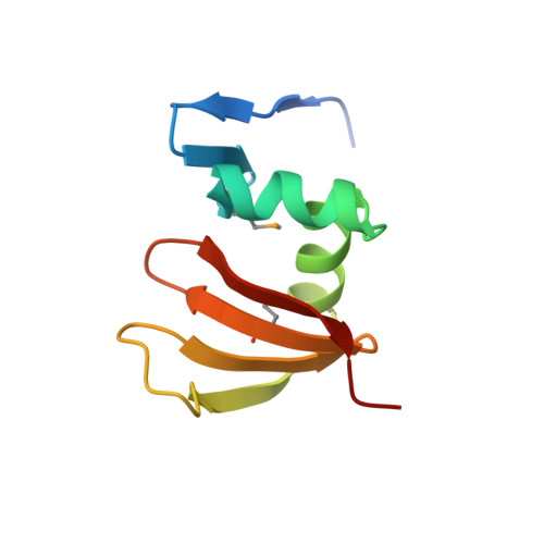
Contact-dependent inhibitor A
A, E
92Escherichia coli NC101Mutation(s): 0 
EC: 3.1
UniProt
Find proteins for P0DSI1 (Escherichia coli (strain NC101))
Explore P0DSI1 
Go to UniProtKB:  P0DSI1
Entity Groups  
Sequence Clusters30% Identity50% Identity70% Identity90% Identity95% Identity100% Identity
UniProt GroupP0DSI1
Sequence Annotations
Expand
  • Reference Sequence
Find similar proteins by:  (by identity cutoff)  |  3D Structure
Entity ID: 2
MoleculeChains Sequence LengthOrganismDetailsImage
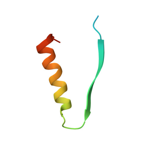
Elongation factor TuB [auth C],
F [auth G]
45Escherichia coli BL21(DE3)Mutation(s): 0 
UniProt
Find proteins for P0CE48 (Escherichia coli (strain K12))
Explore P0CE48 
Go to UniProtKB:  P0CE48
Entity Groups  
Sequence Clusters30% Identity50% Identity70% Identity90% Identity95% Identity100% Identity
UniProt GroupP0CE48
Sequence Annotations
Expand
  • Reference Sequence
Find similar proteins by:  (by identity cutoff)  |  3D Structure
Entity ID: 3
MoleculeChains Sequence LengthOrganismDetailsImage
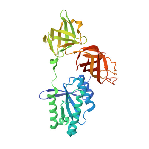
Elongation factor TuC [auth D],
G [auth H]
335Escherichia coli BL21(DE3)Mutation(s): 0 
UniProt
Find proteins for P0CE47 (Escherichia coli (strain K12))
Explore P0CE47 
Go to UniProtKB:  P0CE47
Entity Groups  
Sequence Clusters30% Identity50% Identity70% Identity90% Identity95% Identity100% Identity
UniProt GroupP0CE47
Sequence Annotations
Expand
  • Reference Sequence
Find similar proteins by:  (by identity cutoff)  |  3D Structure
Entity ID: 4
MoleculeChains Sequence LengthOrganismDetailsImage
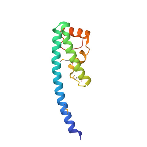
Contact-dependent inhibitor ID [auth B],
H [auth F]
114Escherichia coli NC101Mutation(s): 0 
UniProt
Find proteins for P0DSM8 (Escherichia coli (strain NC101))
Explore P0DSM8 
Go to UniProtKB:  P0DSM8
Entity Groups  
Sequence Clusters30% Identity50% Identity70% Identity90% Identity95% Identity100% Identity
UniProt GroupP0DSM8
Sequence Annotations
Expand
  • Reference Sequence
Small Molecules
Modified Residues  1 Unique
IDChains TypeFormula2D DiagramParent
MSE
Query on MSE
A, E
L-PEPTIDE LINKINGC5 H11 N O2 SeMET
Experimental Data & Validation

Experimental Data

  • Method: X-RAY DIFFRACTION
  • Resolution: 3.30 Å
  • R-Value Free: 0.268 
  • R-Value Work: 0.243 
  • R-Value Observed: 0.244 
  • Space Group: P 1 21 1
Unit Cell:
Length ( Å )Angle ( ˚ )
a = 74.395α = 90
b = 128.354β = 109.58
c = 100.358γ = 90
Software Package:
Software NamePurpose
REFMACrefinement
HKL-3000data reduction
HKL-3000data scaling
PHASERphasing

Structure Validation

View Full Validation Report


Ligand Structure Quality Assessment 


Entry History & Funding Information

Deposition Data


Funding OrganizationLocationGrant Number
National Institutes of Health/National Institute of General Medical Sciences (NIH/NIGMS)United StatesGM094585
National Institutes of Health/National Institute of General Medical Sciences (NIH/NIGMS)United StatesGM102318

Revision History  (Full details and data files)

  • Version 1.0: 2017-06-28
    Type: Initial release
  • Version 1.1: 2017-09-20
    Changes: Author supporting evidence
  • Version 1.2: 2017-10-25
    Changes: Database references
  • Version 1.3: 2019-12-25
    Changes: Author supporting evidence
  • Version 1.4: 2023-09-27
    Changes: Data collection, Database references, Refinement description
  • Version 1.5: 2023-11-15
    Changes: Data collection
  • Version 1.6: 2024-10-30
    Changes: Structure summary