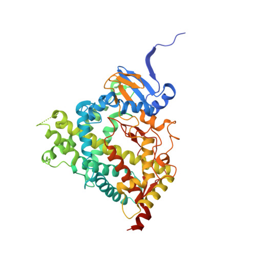
5IRQ

Human cytochrome P450 17A1 bound to inhibitors (R)- and (S)- orteronel


Experimental Data Snapshot

  • Method: X-RAY DIFFRACTION
  • Resolution: 2.20 Å
  • R-Value Free: 0.249 
  • R-Value Work: 0.201 
  • R-Value Observed: 0.204 

Starting Model: experimental
View more details

wwPDB Validation   3D Report Full Report


Ligand Structure Quality Assessment 


This is version 1.5 of the entry. See complete history


Literature

Structural and Functional Evaluation of Clinically Relevant Inhibitors of Steroidogenic Cytochrome P450 17A1.

Petrunak, E.M.Rogers, S.A.Aube, J.Scott, E.E.

(2017) Drug Metab Dispos 45: 635-645

  • DOI: https://doi.org/10.1124/dmd.117.075317
  • Primary Citation of Related Structures:  
    5IRQ, 5IRV

  • PubMed Abstract: 

    Human steroidogenic cytochrome P450 17A1 (CYP17A1) is a bifunctional enzyme that performs both hydroxylation and lyase reactions, with the latter required to generate androgens that fuel prostate cancer proliferation. The steroid abiraterone, the active form of the only CYP17A1 inhibitor approved by the Food and Drug Administration, binds the catalytic heme iron, nonselectively impeding both reactions and ultimately causing undesirable corticosteroid imbalance. Some nonsteroidal inhibitors reportedly inhibit the lyase reaction more than the preceding hydroxylase reaction, which would be clinically advantageous, but the mechanism is not understood. Thus, the nonsteroidal inhibitors seviteronel and orteronel and the steroidal inhibitors abiraterone and galeterone were compared with respect to their binding modes and hydroxylase versus lyase inhibition. Binding studies and X-ray structures of CYP17A1 with nonsteroidal inhibitors reveal coordination to the heme iron like the steroidal inhibitors. ( S )-seviteronel binds similarly to both observed CYP17A1 conformations. However, ( S )-orteronel and ( R )-orteronel bind to distinct CYP17A1 conformations that differ in a region implicated in ligand entry/exit and the presence of a peripheral ligand. To reconcile these binding modes with enzyme function, side-by-side enzymatic analysis was undertaken and revealed that neither the nonsteroidal seviteronel nor the ( S )-orteronel inhibitors demonstrated significant lyase selectivity, but the less potent ( R )-orteronel was 8- to 11-fold selective for lyase inhibition. While active-site iron coordination is consistent with competitive inhibition, conformational selection for binding of some inhibitors and the differential presence of a peripheral ligand molecule suggest the possibility of CYP17A1 functional modulation by features outside the active site.


  • Organizational Affiliation

    Department of Medicinal Chemistry, University of Kansas, Lawrence, Kansas.


Macromolecules
Find similar proteins by:  (by identity cutoff)  |  3D Structure
Entity ID: 1
MoleculeChains Sequence LengthOrganismDetailsImage
Steroid 17-alpha-hydroxylase/17,20 lyase
A, B, C, D
494Homo sapiensMutation(s): 0 
Gene Names: CYP17A1CYP17S17AH
EC: 1.14.14.19 (PDB Primary Data), 4.1.2.30 (PDB Primary Data), 1.14.14.32 (UniProt)
UniProt & NIH Common Fund Data Resources
Find proteins for P05093 (Homo sapiens)
Explore P05093 
Go to UniProtKB:  P05093
PHAROS:  P05093
GTEx:  ENSG00000148795 
Entity Groups  
Sequence Clusters30% Identity50% Identity70% Identity90% Identity95% Identity100% Identity
UniProt GroupP05093
Sequence Annotations
Expand
  • Reference Sequence
Small Molecules
Ligands 3 Unique
IDChains Name / Formula / InChI Key2D Diagram3D Interactions
HEM
Query on HEM

Download Ideal Coordinates CCD File 
E [auth A],
G [auth B],
I [auth C],
L [auth D]
PROTOPORPHYRIN IX CONTAINING FE
C34 H32 Fe N4 O4
KABFMIBPWCXCRK-RGGAHWMASA-L
7D6
Query on 7D6

Download Ideal Coordinates CCD File 
J [auth C],
K [auth D]
(S)-orteronel
C18 H17 N3 O2
OZPFIJIOIVJZMN-SFHVURJKSA-N
6D7
Query on 6D7

Download Ideal Coordinates CCD File 
F [auth A],
H [auth B]
(R)-orteronel
C18 H17 N3 O2
OZPFIJIOIVJZMN-GOSISDBHSA-N
Experimental Data & Validation

Experimental Data

  • Method: X-RAY DIFFRACTION
  • Resolution: 2.20 Å
  • R-Value Free: 0.249 
  • R-Value Work: 0.201 
  • R-Value Observed: 0.204 
  • Space Group: P 21 21 21
Unit Cell:
Length ( Å )Angle ( ˚ )
a = 90.168α = 90
b = 153.204β = 90
c = 168.119γ = 90
Software Package:
Software NamePurpose
PHENIXrefinement
SCALAdata scaling
PHASERphasing
PDB_EXTRACTdata extraction
XDSdata reduction

Structure Validation

View Full Validation Report


Ligand Structure Quality Assessment 


Entry History & Funding Information

Deposition Data


Funding OrganizationLocationGrant Number
National Institutes of Health/National Institute of General Medical Sciences (NIH/NIGMS)United States5R01GM102505

Revision History  (Full details and data files)

  • Version 1.0: 2017-03-15
    Type: Initial release
  • Version 1.1: 2017-04-19
    Changes: Database references
  • Version 1.2: 2017-05-17
    Changes: Database references
  • Version 1.3: 2017-09-13
    Changes: Author supporting evidence
  • Version 1.4: 2019-12-25
    Changes: Author supporting evidence
  • Version 1.5: 2023-09-27
    Changes: Data collection, Database references, Refinement description