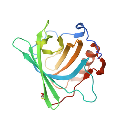
5KHP

Tightening the Recognition of Tetravalent Zr and Th Complexes by the Siderophore-Binding Mammalian Protein Siderocalin for Theranostic Applications


Experimental Data Snapshot

  • Method: X-RAY DIFFRACTION
  • Resolution: 2.65 Å
  • R-Value Free: 0.228 
  • R-Value Work: 0.208 
  • R-Value Observed: 0.209 

Starting Model: experimental
View more details

wwPDB Validation   3D Report Full Report


Ligand Structure Quality Assessment 


This is version 1.3 of the entry. See complete history


Literature

Engineered Recognition of Tetravalent Zirconium and Thorium by Chelator-Protein Systems: Toward Flexible Radiotherapy and Imaging Platforms.

Captain, I.Deblonde, G.J.Rupert, P.B.An, D.D.Illy, M.C.Rostan, E.Ralston, C.Y.Strong, R.K.Abergel, R.J.

(2016) Inorg Chem 55: 11930-11936

  • DOI: https://doi.org/10.1021/acs.inorgchem.6b02041
  • Primary Citation of Related Structures:  
    5KHP, 5KID

  • PubMed Abstract: 

    Targeted α therapy holds tremendous potential as a cancer treatment: it offers the possibility of delivering a highly cytotoxic dose to targeted cells while minimizing damage to surrounding healthy tissue. The metallic α-generating radioisotopes 225 Ac and 227 Th are promising radionuclides for therapeutic use, provided adequate chelation and targeting. Here we demonstrate a new chelating platform composed of a multidentate high-affinity oxygen-donating ligand 3,4,3-LI(CAM) bound to the mammalian protein siderocalin. Respective stability constants log β 110 = 29.65 ± 0.65, 57.26 ± 0.20, and 47.71 ± 0.08, determined for the Eu III (a lanthanide surrogate for Ac III ), Zr IV , and Th IV complexes of 3,4,3-LI(CAM) through spectrophotometric titrations, reveal this ligand to be one of the most powerful chelators for both trivalent and tetravalent metal ions at physiological pH. The resulting metal-ligand complexes are also recognized with extremely high affinity by the siderophore-binding protein siderocalin, with dissociation constants below 40 nM and tight electrostatic interactions, as evidenced by X-ray structures of the protein:ligand:metal adducts with Zr IV and Th IV . Finally, differences in biodistribution profiles between free and siderocalin-bound 238 Pu IV -3,4,3-LI(CAM) complexes confirm in vivo stability of the protein construct. The siderocalin:3,4,3-LI(CAM) assembly can therefore serve as a "lock" to consolidate binding to the therapeutic 225 Ac and 227 Th isotopes or to the positron emission tomography emitter 89 Zr, independent of metal valence state.


  • Organizational Affiliation

    Chemical Sciences Division, Lawrence Berkeley National Laboratory , Berkeley, California 94720, United States.


Macromolecules
Find similar proteins by:  (by identity cutoff)  |  3D Structure
Entity ID: 1
MoleculeChains Sequence LengthOrganismDetailsImage
Neutrophil gelatinase-associated lipocalin
A, B, C
180Homo sapiensMutation(s): 0 
Gene Names: LCN2HNLNGAL
UniProt & NIH Common Fund Data Resources
Find proteins for P80188 (Homo sapiens)
Explore P80188 
Go to UniProtKB:  P80188
PHAROS:  P80188
GTEx:  ENSG00000148346 
Entity Groups  
Sequence Clusters30% Identity50% Identity70% Identity90% Identity95% Identity100% Identity
UniProt GroupP80188
Sequence Annotations
Expand
  • Reference Sequence
Small Molecules
Ligands 4 Unique
IDChains Name / Formula / InChI Key2D Diagram3D Interactions
7K9
Query on 7K9

Download Ideal Coordinates CCD File 
E [auth A],
K [auth B],
M [auth C]
N,N'-(butane-1,4-diyl)bis(N-{3-[(2,3-dihydroxybenzene-1-carbonyl)amino]propyl}-2,3-dihydroxybenzamide)
C38 H42 N4 O12
WNIXXCINEMCWIS-UHFFFAOYSA-N
SO4
Query on SO4

Download Ideal Coordinates CCD File 
F [auth A]
G [auth A]
H [auth A]
I [auth A]
N [auth C]
F [auth A],
G [auth A],
H [auth A],
I [auth A],
N [auth C],
O [auth C],
P [auth C],
Q [auth C]
SULFATE ION
O4 S
QAOWNCQODCNURD-UHFFFAOYSA-L
GOL
Query on GOL

Download Ideal Coordinates CCD File 
R [auth C]GLYCEROL
C3 H8 O3
PEDCQBHIVMGVHV-UHFFFAOYSA-N
ZR
Query on ZR

Download Ideal Coordinates CCD File 
D [auth A],
J [auth B],
L [auth C]
ZIRCONIUM ION
Zr
GBNDTYKAOXLLID-UHFFFAOYSA-N
Experimental Data & Validation

Experimental Data

  • Method: X-RAY DIFFRACTION
  • Resolution: 2.65 Å
  • R-Value Free: 0.228 
  • R-Value Work: 0.208 
  • R-Value Observed: 0.209 
  • Space Group: P 41 21 2
Unit Cell:
Length ( Å )Angle ( ˚ )
a = 114.383α = 90
b = 114.383β = 90
c = 117.121γ = 90
Software Package:
Software NamePurpose
REFMACrefinement
HKL-2000data reduction
SCALEPACKdata scaling
PHASERphasing

Structure Validation

View Full Validation Report


Ligand Structure Quality Assessment 


Entry History 

Deposition Data

Revision History  (Full details and data files)

  • Version 1.0: 2017-04-26
    Type: Initial release
  • Version 1.1: 2017-09-27
    Changes: Database references
  • Version 1.2: 2023-09-27
    Changes: Data collection, Database references, Refinement description
  • Version 1.3: 2024-11-06
    Changes: Structure summary