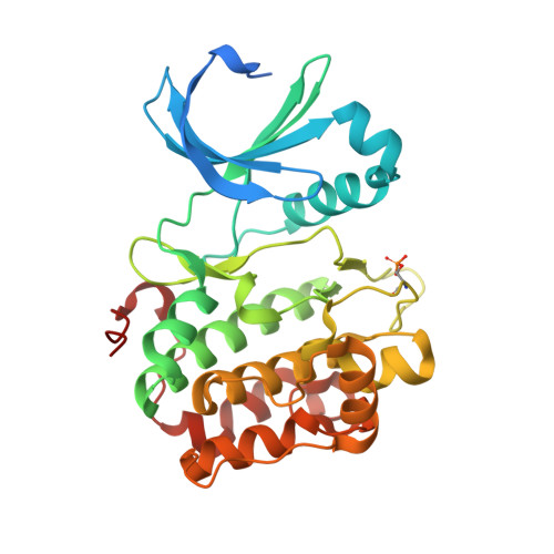
5LVM

Human PDK1 Kinase Domain in Complex with Adenine Bound to the ATP-Binding Site


Experimental Data Snapshot

  • Method: X-RAY DIFFRACTION
  • Resolution: 1.26 Å
  • R-Value Free: 0.155 
  • R-Value Work: 0.134 
  • R-Value Observed: 0.135 

Starting Model: experimental
View more details

wwPDB Validation   3D Report Full Report


Ligand Structure Quality Assessment 


This is version 1.4 of the entry. See complete history


Literature

Bidirectional Allosteric Communication between the ATP-Binding Site and the Regulatory PIF Pocket in PDK1 Protein Kinase.

Schulze, J.O.Saladino, G.Busschots, K.Neimanis, S.Su, E.Odadzic, D.Zeuzem, S.Hindie, V.Herbrand, A.K.Lisa, M.N.Alzari, P.M.Gervasio, F.L.Biondi, R.M.

(2016) Cell Chem Biol 23: 1193-1205

  • DOI: https://doi.org/10.1016/j.chembiol.2016.06.017
  • Primary Citation of Related Structures:  
    5LVL, 5LVM, 5LVN, 5LVO, 5LVP

  • PubMed Abstract: 

    Allostery is a phenomenon observed in many proteins where binding of a macromolecular partner or a small-molecule ligand at one location leads to specific perturbations at a site not in direct contact with the region where the binding occurs. The list of proteins under allosteric regulation includes AGC protein kinases. AGC kinases have a conserved allosteric site, the phosphoinositide-dependent protein kinase 1 (PDK1)-interacting fragment (PIF) pocket, which regulates protein ATP-binding, activity, and interaction with substrates. In this study, we identify small molecules that bind to the ATP-binding site and affect the PIF pocket of AGC kinase family members, PDK1 and Aurora kinase. We describe the mechanistic details and show that although PDK1 and Aurora kinase inhibitors bind to the conserved ATP-binding site, they differentially modulate physiological interactions at the PIF-pocket site. Our work outlines a strategy for developing bidirectional small-molecule allosteric modulators of protein kinases and other signaling proteins.


  • Organizational Affiliation

    Research Group PhosphoSites, Department of Internal Medicine I, Universitätsklinikum Frankfurt, Theodor-Stern-Kai 7, 60590 Frankfurt, Germany.


Macromolecules
Find similar proteins by:  (by identity cutoff)  |  3D Structure
Entity ID: 1
MoleculeChains Sequence LengthOrganismDetailsImage
3-phosphoinositide-dependent protein kinase 1311Homo sapiensMutation(s): 2 
Gene Names: PDPK1PDK1
EC: 2.7.11.1
UniProt & NIH Common Fund Data Resources
Find proteins for O15530 (Homo sapiens)
Explore O15530 
Go to UniProtKB:  O15530
PHAROS:  O15530
GTEx:  ENSG00000140992 
Entity Groups  
Sequence Clusters30% Identity50% Identity70% Identity90% Identity95% Identity100% Identity
UniProt GroupO15530
Sequence Annotations
Expand
  • Reference Sequence
Small Molecules
Modified Residues  1 Unique
IDChains TypeFormula2D DiagramParent
SEP
Query on SEP
A
L-PEPTIDE LINKINGC3 H8 N O6 PSER
Experimental Data & Validation

Experimental Data

  • Method: X-RAY DIFFRACTION
  • Resolution: 1.26 Å
  • R-Value Free: 0.155 
  • R-Value Work: 0.134 
  • R-Value Observed: 0.135 
  • Space Group: C 1 2 1
Unit Cell:
Length ( Å )Angle ( ˚ )
a = 148.02α = 90
b = 44.44β = 102.11
c = 47.78γ = 90
Software Package:
Software NamePurpose
PHENIXrefinement
XDSdata reduction
XSCALEdata scaling
PHASERphasing

Structure Validation

View Full Validation Report


Ligand Structure Quality Assessment 


Entry History & Funding Information

Deposition Data


Funding OrganizationLocationGrant Number
BMBF GO-BioGermany0315102
German Research FoundationGermanyBI 1044/2-3
German Research FoundationGermanyBI 1044/12-1

Revision History  (Full details and data files)

  • Version 1.0: 2016-10-19
    Type: Initial release
  • Version 1.1: 2016-11-02
    Changes: Database references
  • Version 1.2: 2019-10-16
    Changes: Author supporting evidence, Data collection
  • Version 1.3: 2024-01-17
    Changes: Data collection, Database references, Refinement description
  • Version 1.4: 2024-11-20
    Changes: Structure summary