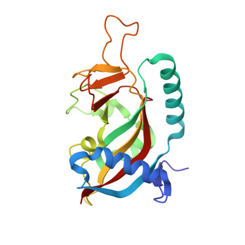
5LX6

Human PARP10 (ARTD10), catalytic fragment in complex with PARP inhibitor Veliparib


Experimental Data Snapshot

  • Method: X-RAY DIFFRACTION
  • Resolution: 1.25 Å
  • R-Value Free: 0.213 
  • R-Value Work: 0.208 
  • R-Value Observed: 0.208 

Starting Model: experimental
View more details

wwPDB Validation   3D Report Full Report


Ligand Structure Quality Assessment 


This is version 1.2 of the entry. See complete history


Literature

Structural Basis for Potency and Promiscuity in Poly(ADP-ribose) Polymerase (PARP) and Tankyrase Inhibitors.

Thorsell, A.G.Ekblad, T.Karlberg, T.Low, M.Pinto, A.F.Tresaugues, L.Moche, M.Cohen, M.S.Schuler, H.

(2017) J Med Chem 60: 1262-1271

  • DOI: https://doi.org/10.1021/acs.jmedchem.6b00990
  • Primary Citation of Related Structures:  
    4R5W, 4R6E, 4RV6, 4TVJ, 4UND, 4UXB, 5LX6

  • PubMed Abstract: 

    Selective inhibitors could help unveil the mechanisms by which inhibition of poly(ADP-ribose) polymerases (PARPs) elicits clinical benefits in cancer therapy. We profiled 10 clinical PARP inhibitors and commonly used research tools for their inhibition of multiple PARP enzymes. We also determined crystal structures of these compounds bound to PARP1 or PARP2. Veliparib and niraparib are selective inhibitors of PARP1 and PARP2; olaparib, rucaparib, and talazoparib are more potent inhibitors of PARP1 but are less selective. PJ34 and UPF1069 are broad PARP inhibitors; PJ34 inserts a flexible moiety into hydrophobic subpockets in various ADP-ribosyltransferases. XAV939 is a promiscuous tankyrase inhibitor and a potent inhibitor of PARP1 in vitro and in cells, whereas IWR1 and AZ-6102 are tankyrase selective. Our biochemical and structural analysis of PARP inhibitor potencies establishes a molecular basis for either selectivity or promiscuity and provides a benchmark for experimental design in assessment of PARP inhibitor effects.


  • Organizational Affiliation

    Program in Chemical Biology and Department of Physiology and Pharmacology, Health & Science University , Portland, Oregon 97210, United States.


Macromolecules
Find similar proteins by:  (by identity cutoff)  |  3D Structure
Entity ID: 1
MoleculeChains Sequence LengthOrganismDetailsImage
Poly [ADP-ribose] polymerase 10
A, B
191Homo sapiensMutation(s): 0 
Gene Names: PARP10
EC: 2.4.2.30 (PDB Primary Data), 2.4.2 (UniProt)
UniProt & NIH Common Fund Data Resources
Find proteins for Q53GL7 (Homo sapiens)
Explore Q53GL7 
Go to UniProtKB:  Q53GL7
PHAROS:  Q53GL7
GTEx:  ENSG00000178685 
Entity Groups  
Sequence Clusters30% Identity50% Identity70% Identity90% Identity95% Identity100% Identity
UniProt GroupQ53GL7
Sequence Annotations
Expand
  • Reference Sequence
Small Molecules
Binding Affinity Annotations 
IDSourceBinding Affinity
78P BindingDB:  5LX6 IC50: min: 1900, max: 4.38e+4 (nM) from 4 assay(s)
Experimental Data & Validation

Experimental Data

  • Method: X-RAY DIFFRACTION
  • Resolution: 1.25 Å
  • R-Value Free: 0.213 
  • R-Value Work: 0.208 
  • R-Value Observed: 0.208 
  • Space Group: P 1 21 1
Unit Cell:
Length ( Å )Angle ( ˚ )
a = 52.48α = 90
b = 88.677β = 104.68
c = 58.405γ = 90
Software Package:
Software NamePurpose
BUSTERrefinement
XDSdata reduction
XSCALEdata scaling
PHASERphasing

Structure Validation

View Full Validation Report


Ligand Structure Quality Assessment 


Entry History 

Deposition Data

Revision History  (Full details and data files)

  • Version 1.0: 2017-01-11
    Type: Initial release
  • Version 1.1: 2017-03-08
    Changes: Database references
  • Version 1.2: 2024-01-17
    Changes: Data collection, Database references, Refinement description, Structure summary