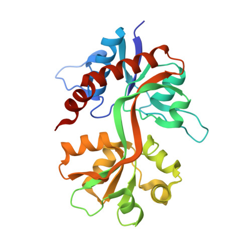
Identification and Structure-Function Study of Positive Allosteric Modulators of Kainate Receptors.
Larsen, A.P., Fievre, S., Frydenvang, K., Francotte, P., Pirotte, B., Kastrup, J.S., Mulle, C.(2017) Mol Pharmacol 91: 576-585
- PubMed: 28360094
- DOI: https://doi.org/10.1124/mol.116.107599
- Primary Citation of Related Structures:
5MFQ, 5MFV, 5MFW - PubMed Abstract:
Kainate receptors (KARs) consist of a class of ionotropic glutamate receptors, which exert diverse pre- and postsynaptic functions through complex signaling regulating the activity of neural circuits. Whereas numerous small-molecule positive allosteric modulators of the ligand-binding domain of ( S )-2-amino-3-(3-hydroxy-5-methylisoxazol-4-yl)propanoic acid (AMPA) receptors have been reported, no such ligands are available for KARs. In this study, we investigated the ability of three benzothiadiazine-based modulators to potentiate glutamate-evoked currents at recombinantly expressed KARs. 4-cyclopropyl-7-fluoro-3,4-dihydro-2 H -1,2,4-benzothiadiazine 1,1-dioxide (BPAM344) potentiated glutamate-evoked currents of GluK2a 21-fold at the highest concentration tested (200 μ M), with an EC 50 of 79 μ M. BPAM344 markedly decreased desensitization kinetics (from 5.5 to 775 ms), whereas it only had a minor effect on deactivation kinetics. 4-cyclopropyl-7-hydroxy-3,4-dihydro-2 H -1,2,4-benzothiadiazine 1,1-dioxide (BPAM521) potentiated the recorded peak current amplitude of GluK2a 12-fold at a concentration of 300 μ M with an EC 50 value of 159 μ M, whereas no potentiation of the glutamate-evoked response was observed for 7-chloro-4-(2-fluoroethyl)-3,4-dihydro-2 H -1,2,4-benzothiadiazine 1,1-dioxide (BPAM121) at the highest concentration of modulator tested (300 μ M). BPAM344 (100 μ M) also potentiated the peak current amplitude of KAR subunits GluK3a (59-fold), GluK2a (15-fold), GluK1b (5-fold), as well as the AMPA receptor subunit GluA1 i (5-fold). X-ray structures of the three modulators in the GluK1 ligand-binding domain were determined, locating two modulator-binding sites at the GluK1 dimer interface. In conclusion, this study may enable the design of new positive allosteric modulators selective for KARs, which will be of great interest for further investigation of the function of KARs in vivo and may prove useful for pharmacologically controlling the activity of neuronal networks.
- Biostructural Research, Department of Drug Design and Pharmacology, Faculty of Health and Medical Sciences, University of Copenhagen, Copenhagen, Denmark (A.P.L., K.F., J.S.K.); Interdisciplinary Institute for Neuroscience, University of Bordeaux, Centre National de la Recherche Scientifique, Unité Mixte de Recherche 5297, Bordeaux, France (A.P.L., S.F., C.M.); and Department of Medicinal Chemistry, Center for Interdisciplinary Research on Medicines, University of Liège, Liège, Belgium (P.F., B.P.).
Organizational Affiliation:


