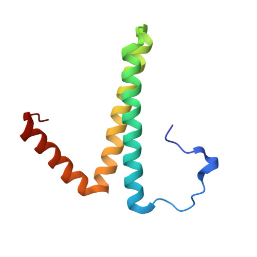
5N5E

Crystal structure of encapsulated ferritin domain from Pyrococcus furiosus PFC_05175


Experimental Data Snapshot

  • Method: X-RAY DIFFRACTION
  • Resolution: 2.03 Å
  • R-Value Free: 0.202 
  • R-Value Work: 0.174 
  • R-Value Observed: 0.165 

Starting Model: experimental
View more details

wwPDB Validation   3D Report Full Report


This is version 1.2 of the entry. See complete history


Literature

Conservation of the structural and functional architecture of encapsulated ferritins in bacteria and archaea.

He, D.Piergentili, C.Ross, J.Tarrant, E.Tuck, L.R.Mackay, C.L.McIver, Z.Waldron, K.J.Clarke, D.J.Marles-Wright, J.

(2019) Biochem J 476: 975-989

  • DOI: https://doi.org/10.1042/BCJ20180922
  • Primary Citation of Related Structures:  
    5N5E, 5N5F

  • PubMed Abstract: 

    Ferritins are a large family of intracellular proteins that protect the cell from oxidative stress by catalytically converting Fe(II) into less toxic Fe(III) and storing iron minerals within their core. Encapsulated ferritins (EncFtn) are a sub-family of ferritin-like proteins, which are widely distributed in all bacterial and archaeal phyla. The recently characterized Rhodospirillum rubrum EncFtn displays an unusual structure when compared with classical ferritins, with an open decameric structure that is enzymatically active, but unable to store iron. This EncFtn must be associated with an encapsulin nanocage in order to act as an iron store. Given the wide distribution of the EncFtn family in organisms with diverse environmental niches, a question arises as to whether this unusual structure is conserved across the family. Here, we characterize EncFtn proteins from the halophile Haliangium ochraceum and the thermophile Pyrococcus furiosus , which show the conserved annular pentamer of dimers topology. Key structural differences are apparent between the homologues, particularly in the centre of the ring and the secondary metal-binding site, which is not conserved across the homologues. Solution and native mass spectrometry analyses highlight that the stability of the protein quaternary structure differs between EncFtn proteins from different species. The ferroxidase activity of EncFtn proteins was confirmed, and we show that while the quaternary structure around the ferroxidase centre is distinct from classical ferritins, the ferroxidase activity is still inhibited by Zn(II). Our results highlight the common structural organization and activity of EncFtn proteins, despite diverse host environments and contexts within encapsulins.


  • Organizational Affiliation

    Institute of Quantitative Biology, Biochemistry, and Biotechnology, School of Biological Sciences, The University of Edinburgh, Max Born Crescent, Edinburgh EH9 3BF, U.K.


Macromolecules
Find similar proteins by:  (by identity cutoff)  |  3D Structure
Entity ID: 1
MoleculeChains Sequence LengthOrganismDetailsImage
PFC_0517599Pyrococcus furiosus COM1Mutation(s): 0 
Gene Names: PFC_05175
EC: 1.16.3.1
UniProt
Find proteins for Q8U1L4 (Pyrococcus furiosus (strain ATCC 43587 / DSM 3638 / JCM 8422 / Vc1))
Explore Q8U1L4 
Go to UniProtKB:  Q8U1L4
Entity Groups  
Sequence Clusters30% Identity50% Identity70% Identity90% Identity95% Identity100% Identity
UniProt GroupQ8U1L4
Sequence Annotations
Expand
  • Reference Sequence
Small Molecules
Ligands 1 Unique
IDChains Name / Formula / InChI Key2D Diagram3D Interactions
FE
Query on FE

Download Ideal Coordinates CCD File 
AB [auth Q]
BB [auth Q]
CB [auth T]
DB [auth T]
EA [auth e]
AB [auth Q],
BB [auth Q],
CB [auth T],
DB [auth T],
EA [auth e],
EB [auth X],
FA [auth e],
FB [auth X],
GA [auth A],
GB [auth a],
HA [auth A],
HB [auth a],
IA [auth B],
JA [auth B],
KA [auth C],
LA [auth C],
MA [auth D],
NA [auth D],
OA [auth E],
PA [auth E],
QA [auth F],
RA [auth F],
SA [auth G],
TA [auth G],
UA [auth L],
VA [auth L],
WA [auth M],
XA [auth M],
YA [auth P],
ZA [auth P]
FE (III) ION
Fe
VTLYFUHAOXGGBS-UHFFFAOYSA-N
Experimental Data & Validation

Experimental Data

  • Method: X-RAY DIFFRACTION
  • Resolution: 2.03 Å
  • R-Value Free: 0.202 
  • R-Value Work: 0.174 
  • R-Value Observed: 0.165 
  • Space Group: P 1 21 1
Unit Cell:
Length ( Å )Angle ( ˚ )
a = 99.853α = 90
b = 110.057β = 91.22
c = 136.271γ = 90
Software Package:
Software NamePurpose
PHENIXrefinement
Aimlessdata scaling
XDSdata reduction
PHASERphasing

Structure Validation

View Full Validation Report


Entry History & Funding Information

Deposition Data


Funding OrganizationLocationGrant Number
Biotechnology and Biological Sciences Research CouncilUnited KingdomBB/N005570/1
China Scholarships CouncilChina--

Revision History  (Full details and data files)

  • Version 1.0: 2018-01-24
    Type: Initial release
  • Version 1.1: 2019-08-07
    Changes: Data collection, Database references
  • Version 1.2: 2024-01-17
    Changes: Data collection, Database references, Derived calculations, Refinement description