5NU6 | pdb_00005nu6

Structure of non-fluorescent human amniotic fluid RBP4


Experimental Data Snapshot

  • Method: X-RAY DIFFRACTION
  • Resolution: 1.68 Å
  • R-Value Free: 
    0.197 (Depositor), 0.221 (DCC) 
  • R-Value Work: 
    0.169 (Depositor), 0.218 (DCC) 
  • R-Value Observed: 
    0.171 (Depositor) 

Starting Model: experimental
View more details

wwPDB Validation 3D Report Full Report

Validation slider image for 5NU6

Ligand Structure Quality Assessment 


This is version 1.2 of the entry. See complete history

Literature

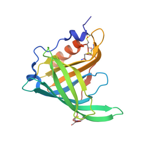
Human plasma retinol-binding protein (RBP4) is also a fatty acid-binding protein.

Perduca, M.Nicolis, S.Mannucci, B.Galliano, M.Monaco, H.L.

(2018) Biochim Biophys Acta 1863: 458-466

  • DOI: https://doi.org/10.1016/j.bbalip.2018.01.010
  • Primary Citation Related Structures: 
    5NTY, 5NU2, 5NU6, 5NU7, 5NU8, 5NU9, 5NUA, 5NUB

  • PubMed Abstract: 

    RBP4 (plasma retinol-binding protein) is the 21 kDa transporter of all-trans retinol that circulates in plasma as a moderately tight 1:1 molar complex of the vitamin with the protein. RBP4 is primarily synthesized in the liver but is also produced by adipose tissue and circulates bound to a larger protein, transthyretin, TTR, that serves to increase its molecular mass and thus avoid its elimination by glomerular filtration. This paper reports the high resolution three-dimensional structures of human RBP4 naturally lacking bound retinol purified from plasma, urine and amniotic fluid. In all these crystals we found a fatty acid molecule bound in the hydrophobic ligand-binding site, a result confirmed by mass spectrometry measurements. In addition we also report the 1.5 Å resolution structures of human holo-RBP4 and of the protein saturated with palmitic and lauric acid and discuss the interaction of the fatty acids and retinol with the protein.


  • Organizational Affiliation
    • Biocrystallography Laboratory, Department of Biotechnology, University of Verona, Ca Vignal 1, strada Le Grazie 15, 37134 Verona, Italy.

Macromolecule Content 

  • Total Structure Weight: 21.31 kDa 
  • Atom Count: 1,517 
  • Modeled Residue Count: 175 
  • Deposited Residue Count: 182 
  • Unique protein chains: 1

Macromolecules

Find similar proteins by:|  3D Structure
Entity ID: 1
MoleculeChains  Sequence LengthOrganismDetailsImage
Retinol-binding protein 4182Homo sapiensMutation(s): 0 
UniProt & NIH Common Fund Data Resources
Find proteins for P02753 (Homo sapiens)
Explore P02753 
Go to UniProtKB:  P02753
PHAROS:  P02753
GTEx:  ENSG00000138207 
Entity Groups
Sequence Clusters30% Identity50% Identity70% Identity90% Identity95% Identity100% Identity
UniProt GroupP02753
Sequence Annotations
Expand
Reference Sequence

Experimental Data & Validation

Experimental Data

  • Method: X-RAY DIFFRACTION
  • Resolution: 1.68 Å
  • R-Value Free:  0.197 (Depositor), 0.221 (DCC) 
  • R-Value Work:  0.169 (Depositor), 0.218 (DCC) 
  • R-Value Observed: 0.171 (Depositor) 
Space Group: H 3
Unit Cell:
Length ( Å )Angle ( ˚ )
a = 102.58α = 90
b = 102.58β = 90
c = 73.282γ = 120
Software Package:
Software NamePurpose
REFMACrefinement
iMOSFLMdata reduction
SCALAdata scaling

Structure Validation

View Full Validation Report


Ligand Structure Quality Assessment 


Entry History 

Deposition Data

Revision History  (Full details and data files)

  • Version 1.0: 2018-03-07
    Type: Initial release
  • Version 1.1: 2024-01-17
    Changes: Data collection, Database references, Refinement description
  • Version 1.2: 2024-10-23
    Changes: Structure summary