5TX6 | pdb_00005tx6

Structure of TGF-beta2 derivative with deletion of residues 52-71 and 10 single amino acid mutations (mmTGF-beta2-7M)

  • Classification: CYTOKINE
  • Organism(s): Mus musculus
  • Expression System: Escherichia coli
  • Mutation(s): Yes 

  • Deposited: 2016-11-15 Released: 2017-03-01 
  • Deposition Author(s): Petrunak, E.M., Hinck, A.P.
  • Funding Organization(s): National Institutes of Health/National Institute of General Medical Sciences (NIH/NIGMS), National Institutes of Health/National Cancer Institute (NIH/NCI), Robert A. Welch Foundation

Experimental Data Snapshot

  • Method: X-RAY DIFFRACTION
  • Resolution: 2.75 Å
  • R-Value Free: 
    0.272 (Depositor), 0.272 (DCC) 
  • R-Value Work: 
    0.213 (Depositor), 0.217 (DCC) 
  • R-Value Observed: 
    0.216 (Depositor) 

Starting Model: experimental
View more details

wwPDB Validation 3D Report Full Report

Validation slider image for 5TX6

This is version 1.5 of the entry. See complete history

Literature

An engineered transforming growth factor beta (TGF-beta ) monomer that functions as a dominant negative to block TGF-beta signaling.

Kim, S.K.Barron, L.Hinck, C.S.Petrunak, E.M.Cano, K.E.Thangirala, A.Iskra, B.Brothers, M.Vonberg, M.Leal, B.Richter, B.Kodali, R.Taylor, A.B.Du, S.Barnes, C.O.Sulea, T.Calero, G.Hart, P.J.Hart, M.J.Demeler, B.Hinck, A.P.

(2017) J Biological Chem 292: 7173-7188

  • DOI: https://doi.org/10.1074/jbc.M116.768754
  • Primary Citation Related Structures: 
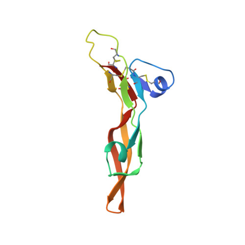
    5TX2, 5TX4, 5TX6

  • PubMed Abstract: 

    The transforming growth factor β isoforms, TGF-β1, -β2, and -β3, are small secreted homodimeric signaling proteins with essential roles in regulating the adaptive immune system and maintaining the extracellular matrix. However, dysregulation of the TGF-β pathway is responsible for promoting the progression of several human diseases, including cancer and fibrosis. Despite the known importance of TGF-βs in promoting disease progression, no inhibitors have been approved for use in humans. Herein, we describe an engineered TGF-β monomer, lacking the heel helix, a structural motif essential for binding the TGF-β type I receptor (TβRI) but dispensable for binding the other receptor required for TGF-β signaling, the TGF-β type II receptor (TβRII), as an alternative therapeutic modality for blocking TGF-β signaling in humans. As shown through binding studies and crystallography, the engineered monomer retained the same overall structure of native TGF-β monomers and bound TβRII in an identical manner. Cell-based luciferase assays showed that the engineered monomer functioned as a dominant negative to inhibit TGF-β signaling with a K i of 20-70 nm Investigation of the mechanism showed that the high affinity of the engineered monomer for TβRII, coupled with its reduced ability to non-covalently dimerize and its inability to bind and recruit TβRI, enabled it to bind endogenous TβRII but prevented it from binding and recruiting TβRI to form a signaling complex. Such engineered monomers provide a new avenue to probe and manipulate TGF-β signaling and may inform similar modifications of other TGF-β family members.


  • Organizational Affiliation
    • the Departments of Biochemistry and Structural Biology and.

Macromolecule Content 

  • Total Structure Weight: 31.94 kDa 
  • Atom Count: 2,150 
  • Modeled Residue Count: 261 
  • Deposited Residue Count: 279 
  • Unique protein chains: 1

Macromolecules

Find similar proteins by:|  3D Structure
Entity ID: 1
MoleculeChains  Sequence LengthOrganismDetailsImage
Transforming growth factor beta-2A [auth B],
B [auth A],
C
93Mus musculusMutation(s): 10 
Gene Names: Tgfb2
UniProt
Find proteins for P27090 (Mus musculus)
Explore P27090 
Go to UniProtKB:  P27090
Entity Groups
Sequence Clusters30% Identity50% Identity70% Identity90% Identity95% Identity100% Identity
UniProt GroupP27090
Sequence Annotations
Expand
Reference Sequence

Small Molecules

Ligands 1 Unique
IDChains Name / Formula / InChI Key2D Diagram3D Interactions
CA

Query on CA


Download:Ideal Coordinates CCD File
D [auth A]CALCIUM ION
Ca
BHPQYMZQTOCNFJ-UHFFFAOYSA-N

Experimental Data & Validation

Experimental Data

  • Method: X-RAY DIFFRACTION
  • Resolution: 2.75 Å
  • R-Value Free:  0.272 (Depositor), 0.272 (DCC) 
  • R-Value Work:  0.213 (Depositor), 0.217 (DCC) 
  • R-Value Observed: 0.216 (Depositor) 
Space Group: P 31 2 1
Unit Cell:
Length ( Å )Angle ( ˚ )
a = 81.744α = 90
b = 81.744β = 90
c = 80.926γ = 120
Software Package:
Software NamePurpose
PHENIXrefinement
iMOSFLMdata reduction
SCALAdata scaling
PHASERphasing

Structure Validation

View Full Validation Report


Entry History 

& Funding Information

Deposition Data


Funding OrganizationLocationGrant Number
National Institutes of Health/National Institute of General Medical Sciences (NIH/NIGMS)United StatesGM58670
National Institutes of Health/National Cancer Institute (NIH/NCI)United StatesCA172886
Robert A. Welch FoundationUnited StatesAQ1842

Revision History  (Full details and data files)

  • Version 1.0: 2017-03-01
    Type: Initial release
  • Version 1.1: 2017-03-08
    Changes: Database references
  • Version 1.2: 2017-05-10
    Changes: Database references
  • Version 1.3: 2017-09-27
    Changes: Author supporting evidence
  • Version 1.4: 2019-12-04
    Changes: Author supporting evidence
  • Version 1.5: 2024-10-09
    Changes: Data collection, Database references, Derived calculations, Refinement description, Structure summary