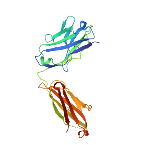
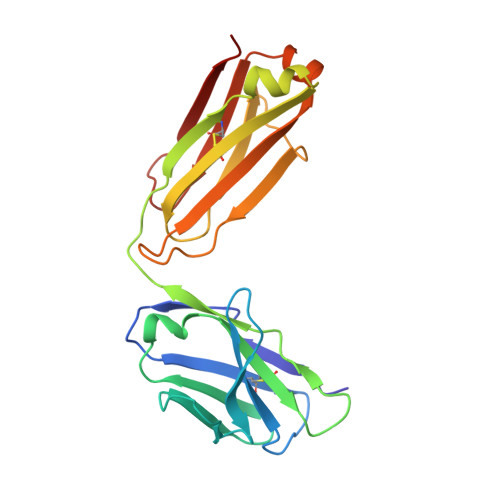
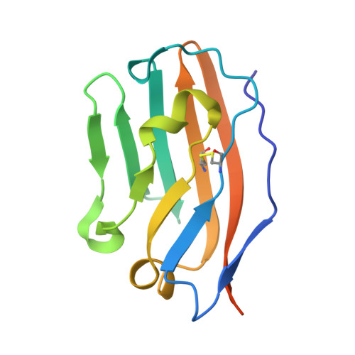
5TZ2

Crystal structure of human CD47 ECD bound to Fab of C47B222


Experimental Data Snapshot

  • Method: X-RAY DIFFRACTION
  • Resolution: 2.30 Å
  • R-Value Free: 0.255 
  • R-Value Work: 0.201 
  • R-Value Observed: 0.204 

Starting Model: experimental
View more details

wwPDB Validation   3D Report Full Report


Ligand Structure Quality Assessment 


This is version 2.2 of the entry. See complete history


Literature

Anti-leukemic activity and tolerability of anti-human CD47 monoclonal antibodies.

Pietsch, E.C.Dong, J.Cardoso, R.Zhang, X.Chin, D.Hawkins, R.Dinh, T.Zhou, M.Strake, B.Feng, P.H.Rocca, M.Santos, C.D.Shan, X.Danet-Desnoyers, G.Shi, F.Kaiser, E.Millar, H.J.Fenton, S.Swanson, R.Nemeth, J.A.Attar, R.M.

(2017) Blood Cancer J 7: e536-e536

  • DOI: https://doi.org/10.1038/bcj.2017.7
  • Primary Citation of Related Structures:  
    5TZ2, 5TZT, 5TZU

  • PubMed Abstract: 

    CD47, a broadly expressed cell surface protein, inhibits cell phagocytosis via interaction with phagocyte-expressed SIRPα. A variety of hematological malignancies demonstrate elevated CD47 expression, suggesting that CD47 may mediate immune escape. We discovered three unique CD47-SIRPα blocking anti-CD47 monoclonal antibodies (mAbs) with low nano-molar affinity to human and cynomolgus monkey CD47, and no hemagglutination and platelet aggregation activity. To characterize the anti-cancer activity elicited by blocking CD47, the mAbs were cloned into effector function silent and competent Fc backbones. Effector function competent mAbs demonstrated potent activity in vitro and in vivo, while effector function silent mAbs demonstrated minimal activity, indicating that blocking CD47 only leads to a therapeutic effect in the presence of Fc effector function. A non-human primate study revealed that the effector function competent mAb IgG1 C47B222-(CHO) decreased red blood cells (RBC), hematocrit and hemoglobin by >40% at 1 mg/kg, whereas the effector function silent mAb IgG2σ C47B222-(CHO) had minimal impact on RBC indices at 1 and 10 mg/kg. Taken together, our findings suggest that targeting CD47 is an attractive therapeutic anti-cancer approach. However, the anti-cancer activity observed with anti-CD47 mAbs is Fc effector dependent as are the side effects observed on RBC indices.


  • Organizational Affiliation

    Oncology Discovery, Janssen Research and Development, Spring House, PA, USA.


Macromolecules
Find similar proteins by:  (by identity cutoff)  |  3D Structure
Entity ID: 1
MoleculeChains Sequence LengthOrganismDetailsImage
C47B222 Fab Heavy ChainA [auth H]229Homo sapiensMutation(s): 0 
Entity Groups  
Sequence Clusters30% Identity50% Identity70% Identity90% Identity95% Identity100% Identity
Sequence Annotations
Expand
  • Reference Sequence
Find similar proteins by:  (by identity cutoff)  |  3D Structure
Entity ID: 2
MoleculeChains Sequence LengthOrganismDetailsImage
C47B222 Fab Light ChainB [auth L]214Homo sapiensMutation(s): 0 
Entity Groups  
Sequence Clusters30% Identity50% Identity70% Identity90% Identity95% Identity100% Identity
Sequence Annotations
Expand
  • Reference Sequence
Find similar proteins by:  (by identity cutoff)  |  3D Structure
Entity ID: 3
MoleculeChains Sequence LengthOrganismDetailsImage
Leukocyte surface antigen CD47129Homo sapiensMutation(s): 1 
Gene Names: CD47MER6
UniProt & NIH Common Fund Data Resources
Find proteins for Q08722 (Homo sapiens)
Explore Q08722 
Go to UniProtKB:  Q08722
PHAROS:  Q08722
GTEx:  ENSG00000196776 
Entity Groups  
Sequence Clusters30% Identity50% Identity70% Identity90% Identity95% Identity100% Identity
UniProt GroupQ08722
Glycosylation
Glycosylation Sites: 5Go to GlyGen: Q08722-1
Sequence Annotations
Expand
  • Reference Sequence
Oligosaccharides

Help

Entity ID: 4
MoleculeChains Length2D Diagram Glycosylation3D Interactions
2-acetamido-2-deoxy-beta-D-glucopyranose-(1-4)-2-acetamido-2-deoxy-beta-D-glucopyranoseD [auth A],
E [auth B]
2N-Glycosylation
Glycosylation Resources
GlyTouCan:  G42666HT
GlyCosmos:  G42666HT
GlyGen:  G42666HT
Experimental Data & Validation

Experimental Data

  • Method: X-RAY DIFFRACTION
  • Resolution: 2.30 Å
  • R-Value Free: 0.255 
  • R-Value Work: 0.201 
  • R-Value Observed: 0.204 
  • Space Group: P 1 21 1
Unit Cell:
Length ( Å )Angle ( ˚ )
a = 60.567α = 90
b = 72.934β = 108.97
c = 72.698γ = 90
Software Package:
Software NamePurpose
HKL-2000data reduction
PHASERphasing
PHENIXrefinement
PDB_EXTRACTdata extraction
HKL-2000data scaling
DENZOdata reduction
SCALEPACKdata scaling

Structure Validation

View Full Validation Report


Ligand Structure Quality Assessment 


Entry History 

Deposition Data

Revision History  (Full details and data files)

  • Version 1.0: 2017-03-15
    Type: Initial release
  • Version 1.1: 2017-11-22
    Changes: Refinement description
  • Version 2.0: 2020-07-29
    Type: Remediation
    Reason: Carbohydrate remediation
    Changes: Advisory, Atomic model, Data collection, Derived calculations, Structure summary
  • Version 2.1: 2023-10-04
    Changes: Data collection, Database references, Refinement description, Structure summary
  • Version 2.2: 2024-11-13
    Changes: Structure summary