5U6R | pdb_00005u6r

E. coli CTP synthase CC mutant filament (product-bound)


Experimental Data Snapshot

  • Method: ELECTRON MICROSCOPY
  • Resolution: 5.70 Å
  • Aggregation State: FILAMENT 
  • Reconstruction Method: HELICAL 

wwPDB Validation 3D Report Full Report

Validation slider image for 5U6R

This is version 1.4 of the entry. See complete history

Literature

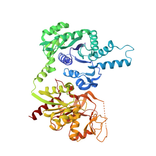
Human CTP synthase filament structure reveals the active enzyme conformation.

Lynch, E.M.Hicks, D.R.Shepherd, M.Endrizzi, J.A.Maker, A.Hansen, J.M.Barry, R.M.Gitai, Z.Baldwin, E.P.Kollman, J.M.

(2017) Nat Struct Mol Biol 24: 507-514

  • DOI: https://doi.org/10.1038/nsmb.3407
  • Primary Citation Related Structures: 
    5TKV, 5U03, 5U05, 5U3C, 5U6R

  • PubMed Abstract: 

    The universally conserved enzyme CTP synthase (CTPS) forms filaments in bacteria and eukaryotes. In bacteria, polymerization inhibits CTPS activity and is required for nucleotide homeostasis. Here we show that for human CTPS, polymerization increases catalytic activity. The cryo-EM structures of bacterial and human CTPS filaments differ considerably in overall architecture and in the conformation of the CTPS protomer, explaining the divergent consequences of polymerization on activity. The structure of human CTPS filament, the first structure of the full-length human enzyme, reveals a novel active conformation. The filament structures elucidate allosteric mechanisms of assembly and regulation that rely on a conserved conformational equilibrium. The findings may provide a mechanism for increasing human CTPS activity in response to metabolic state and challenge the assumption that metabolic filaments are generally storage forms of inactive enzymes. Allosteric regulation of CTPS polymerization by ligands likely represents a fundamental mechanism underlying assembly of other metabolic filaments.


  • Organizational Affiliation
    • Department of Biochemistry, University of Washington, Seattle, Washington, USA.

Macromolecule Content 

  • Total Structure Weight: 245.43 kDa 
  • Atom Count: 16,868 
  • Modeled Residue Count: 2,136 
  • Deposited Residue Count: 2,180 
  • Unique protein chains: 1

Macromolecules

Find similar proteins by:|  3D Structure
Entity ID: 1
MoleculeChains  Sequence LengthOrganismDetailsImage
CTP synthase
A, B, C, D
545Escherichia coliMutation(s): 0 
Gene Names: pyrGECS88_3048
EC: 6.3.4.2
UniProt
Find proteins for P0A7E5 (Escherichia coli (strain K12))
Explore P0A7E5 
Go to UniProtKB:  P0A7E5
Entity Groups
Sequence Clusters30% Identity50% Identity70% Identity90% Identity95% Identity100% Identity
UniProt GroupP0A7E5
Sequence Annotations
Expand
Reference Sequence

Small Molecules

Ligands 2 Unique
IDChains Name / Formula / InChI Key2D Diagram3D Interactions
CTP

Query on CTP


Download:Ideal Coordinates CCD File
E [auth A],
G [auth B],
I [auth C],
K [auth D]
CYTIDINE-5'-TRIPHOSPHATE
C9 H16 N3 O14 P3
PCDQPRRSZKQHHS-XVFCMESISA-N
ADP

Query on ADP


Download:Ideal Coordinates CCD File
F [auth A],
H [auth B],
J [auth C],
L [auth D]
ADENOSINE-5'-DIPHOSPHATE
C10 H15 N5 O10 P2
XTWYTFMLZFPYCI-KQYNXXCUSA-N

Experimental Data & Validation

Experimental Data

  • Method: ELECTRON MICROSCOPY
  • Resolution: 5.70 Å
  • Aggregation State: FILAMENT 
  • Reconstruction Method: HELICAL 
EM Software:
TaskSoftware PackageVersion
RECONSTRUCTIONSPIDER
RECONSTRUCTIONhsearch_lorentz

Structure Validation

View Full Validation Report


Entry History 

Deposition Data

Revision History  (Full details and data files)

  • Version 1.0: 2017-04-26
    Type: Initial release
  • Version 1.1: 2017-05-17
    Changes: Database references
  • Version 1.2: 2017-06-21
    Changes: Database references
  • Version 1.3: 2018-07-18
    Changes: Data collection
  • Version 1.4: 2024-10-09
    Changes: Data collection, Database references, Derived calculations, Structure summary