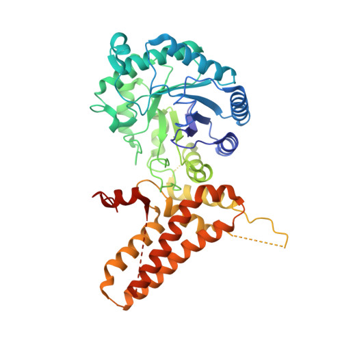
5UN9

The crystal structure of human O-GlcNAcase in complex with Thiamet-G


Experimental Data Snapshot

  • Method: X-RAY DIFFRACTION
  • Resolution: 2.50 Å
  • R-Value Free: 0.260 
  • R-Value Work: 0.210 
  • R-Value Observed: 0.213 

Starting Model: experimental
View more details

wwPDB Validation   3D Report Full Report


Ligand Structure Quality Assessment 


This is version 1.3 of the entry. See complete history


Literature

Structures of human O-GlcNAcase and its complexes reveal a new substrate recognition mode.

Li, B.Li, H.Lu, L.Jiang, J.

(2017) Nat Struct Mol Biol 24: 362-369

  • DOI: https://doi.org/10.1038/nsmb.3390
  • Primary Citation of Related Structures:  
    5TKE, 5UN8, 5UN9

  • PubMed Abstract: 

    Human O-GlcNAcase (hOGA) is the unique enzyme responsible for the hydrolysis of the O-linked β-N-acetyl glucosamine (O-GlcNAc) modification, an essential protein glycosylation event that modulates the function of numerous cellular proteins in response to nutrients and stress. Here we report crystal structures of a truncated hOGA, which comprises the catalytic and stalk domains, in apo form, in complex with an inhibitor, and in complex with a glycopeptide substrate. We found that hOGA forms an unusual arm-in-arm homodimer in which the catalytic domain of one monomer is covered by the stalk domain of the sister monomer to create a substrate-binding cleft. Notably, the residues on the cleft surface afford extensive interactions with the peptide substrate in a recognition mode that is distinct from that of its bacterial homologs. These structures represent the first model of eukaryotic enzymes in the glycoside hydrolase 84 (GH84) family and provide a crucial starting point for understanding the substrate specificity of hOGA, which regulates a broad range of biological and pathological processes.


  • Organizational Affiliation

    Pharmaceutical Sciences Division, School of Pharmacy, University of Wisconsin-Madison, Madison, Wisconsin, USA.


Macromolecules
Find similar proteins by:  (by identity cutoff)  |  3D Structure
Entity ID: 1
MoleculeChains Sequence LengthOrganismDetailsImage
Protein O-GlcNAcase
A, B
504Homo sapiensMutation(s): 0 
Gene Names: MGEA5HEXCKIAA0679MEA5
EC: 3.2.1.169 (PDB Primary Data), 3.2.1 (PDB Primary Data)
UniProt & NIH Common Fund Data Resources
Find proteins for O60502 (Homo sapiens)
Explore O60502 
Go to UniProtKB:  O60502
PHAROS:  O60502
GTEx:  ENSG00000198408 
Entity Groups  
Sequence Clusters30% Identity50% Identity70% Identity90% Identity95% Identity100% Identity
UniProt GroupO60502
Sequence Annotations
Expand
  • Reference Sequence
Small Molecules
Ligands 1 Unique
IDChains Name / Formula / InChI Key2D Diagram3D Interactions
NHT
Query on NHT

Download Ideal Coordinates CCD File 
C [auth A],
D [auth B]
(3AR,5R,6S,7R,7AR)-2-(ETHYLAMINO)-5-(HYDROXYMETHYL)-5,6,7,7A-TETRAHYDRO-3AH-PYRANO[3,2-D][1,3]THIAZOLE-6,7-DIOL
C9 H16 N2 O4 S
PPAIMZHKIXDJRN-FMDGEEDCSA-N
Experimental Data & Validation

Experimental Data

  • Method: X-RAY DIFFRACTION
  • Resolution: 2.50 Å
  • R-Value Free: 0.260 
  • R-Value Work: 0.210 
  • R-Value Observed: 0.213 
  • Space Group: P 1 21 1
Unit Cell:
Length ( Å )Angle ( ˚ )
a = 82.572α = 90
b = 96.106β = 115.01
c = 89.534γ = 90
Software Package:
Software NamePurpose
PHENIXrefinement
HKL-2000data reduction
HKL-2000data scaling
PHASERphasing

Structure Validation

View Full Validation Report


Ligand Structure Quality Assessment 


Entry History & Funding Information

Deposition Data


Funding OrganizationLocationGrant Number
University of Wisconsin-MadisonUnited States--

Revision History  (Full details and data files)

  • Version 1.0: 2017-03-15
    Type: Initial release
  • Version 1.1: 2017-04-05
    Changes: Database references
  • Version 1.2: 2017-04-19
    Changes: Database references
  • Version 1.3: 2023-10-04
    Changes: Data collection, Database references, Refinement description