5UQ9 | pdb_00005uq9

Crystal structure of 6-phosphogluconate dehydrogenase with ((4R,5R)-5-(hydroxycarbamoyl)-2,2-dimethyl-1,3-dioxolan-4-yl)methyl dihydrogen phosphate


Experimental Data Snapshot

  • Method: X-RAY DIFFRACTION
  • Resolution: 3.00 Å
  • R-Value Free: 
    0.272 (Depositor), 0.267 (DCC) 
  • R-Value Work: 
    0.217 (Depositor), 0.216 (DCC) 
  • R-Value Observed: 
    0.220 (Depositor) 

Starting Model: experimental
View more details

wwPDB Validation 3D Report Full Report

Validation slider image for 5UQ9

Ligand Structure Quality Assessment 


This is version 1.2 of the entry. See complete history

Literature

Functional Genomics Reveals Synthetic Lethality between Phosphogluconate Dehydrogenase and Oxidative Phosphorylation.

Sun, Y.Bandi, M.Lofton, T.Smith, M.Bristow, C.A.Carugo, A.Rogers, N.Leonard, P.Chang, Q.Mullinax, R.Han, J.Shi, X.Seth, S.Meyers, B.A.Miller, M.Miao, L.Ma, X.Feng, N.Giuliani, V.Geck Do, M.Czako, B.Palmer, W.S.Mseeh, F.Asara, J.M.Jiang, Y.Morlacchi, P.Zhao, S.Peoples, M.Tieu, T.N.Warmoes, M.O.Lorenzi, P.L.Muller, F.L.DePinho, R.A.Draetta, G.F.Toniatti, C.Jones, P.Heffernan, T.P.Marszalek, J.R.

(2019) Cell Rep 26: 469-482.e5

  • DOI: https://doi.org/10.1016/j.celrep.2018.12.043
  • Primary Citation Related Structures: 
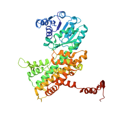
    5UQ9

  • PubMed Abstract: 

    The plasticity of a preexisting regulatory circuit compromises the effectiveness of targeted therapies, and leveraging genetic vulnerabilities in cancer cells may overcome such adaptations. Hereditary leiomyomatosis renal cell carcinoma (HLRCC) is characterized by oxidative phosphorylation (OXPHOS) deficiency caused by fumarate hydratase (FH) nullizyogosity. To identify metabolic genes that are synthetically lethal with OXPHOS deficiency, we conducted a genetic loss-of-function screen and found that phosphogluconate dehydrogenase (PGD) inhibition robustly blocks the proliferation of FH mutant cancer cells both in vitro and in vivo. Mechanistically, PGD inhibition blocks glycolysis, suppresses reductive carboxylation of glutamine, and increases the NADP + /NADPH ratio to disrupt redox homeostasis. Furthermore, in the OXPHOS-proficient context, blocking OXPHOS using the small-molecule inhibitor IACS-010759 enhances sensitivity to PGD inhibition in vitro and in vivo. Together, our study reveals a dependency on PGD in OXPHOS-deficient tumors that might inform therapeutic intervention in specific patient populations.


  • Organizational Affiliation
    • Institute for Applied Cancer Science, The University of Texas MD Anderson Cancer Center, Houston, TX 77030, USA; Center for Co-Clinical Trials, The University of Texas MD Anderson Cancer Center, Houston, TX 77030, USA. Electronic address: ysun8@mdanderson.org.

Macromolecule Content 

  • Total Structure Weight: 430.38 kDa 
  • Atom Count: 29,092 
  • Modeled Residue Count: 3,746 
  • Deposited Residue Count: 3,912 
  • Unique protein chains: 1

Macromolecules

Find similar proteins by:|  3D Structure
Entity ID: 1
MoleculeChains  Sequence LengthOrganismDetailsImage
6-phosphogluconate dehydrogenase, decarboxylating
A, B, C, D, E
A, B, C, D, E, F, G, H
489Homo sapiensMutation(s): 0 
Gene Names: PGDPGDH
EC: 1.1.1.44
UniProt & NIH Common Fund Data Resources
Find proteins for P52209 (Homo sapiens)
Explore P52209 
Go to UniProtKB:  P52209
PHAROS:  P52209
GTEx:  ENSG00000142657 
Entity Groups
Sequence Clusters30% Identity50% Identity70% Identity90% Identity95% Identity100% Identity
UniProt GroupP52209
Sequence Annotations
Expand
Reference Sequence

Small Molecules

Ligands 1 Unique
IDChains Name / Formula / InChI Key2D Diagram3D Interactions
8HS

Query on 8HS


Download:Ideal Coordinates CCD File
I [auth F][(4R,5R)-5-(hydroxycarbamoyl)-2,2-dimethyl-1,3-dioxolan-4-yl]methyl dihydrogen phosphate
C7 H14 N O8 P
DVFAQEVWJBLFDS-RFZPGFLSSA-N
Binding Affinity Annotations 
IDSourceBinding Affinity
8HS BindingDB:  5UQ9 Ki: min: 35, max: 1100 (nM) from 3 assay(s)

Experimental Data & Validation

Experimental Data

  • Method: X-RAY DIFFRACTION
  • Resolution: 3.00 Å
  • R-Value Free:  0.272 (Depositor), 0.267 (DCC) 
  • R-Value Work:  0.217 (Depositor), 0.216 (DCC) 
  • R-Value Observed: 0.220 (Depositor) 
Space Group: P 1 21 1
Unit Cell:
Length ( Å )Angle ( ˚ )
a = 87.311α = 90
b = 164.93β = 91.43
c = 133.199γ = 90
Software Package:
Software NamePurpose
Aimlessdata scaling
PHASERphasing
PHENIXrefinement
PDB_EXTRACTdata extraction
iMOSFLMdata reduction

Structure Validation

View Full Validation Report


Ligand Structure Quality Assessment 


Entry History 

Deposition Data

Revision History  (Full details and data files)

  • Version 1.0: 2018-08-22
    Type: Initial release
  • Version 1.1: 2019-01-23
    Changes: Data collection, Database references
  • Version 1.2: 2023-10-04
    Changes: Data collection, Database references, Refinement description