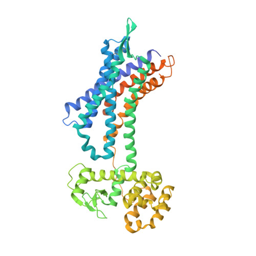
5X33

Leukotriene B4 receptor BLT1 in complex with BIIL260


Experimental Data Snapshot

  • Method: X-RAY DIFFRACTION
  • Resolution: 3.70 Å
  • R-Value Free: 0.296 
  • R-Value Work: 0.248 
  • R-Value Observed: 0.251 

Starting Models: experimental
View more details

wwPDB Validation   3D Report Full Report


Ligand Structure Quality Assessment 


This is version 1.4 of the entry. See complete history


Literature

Na+-mimicking ligands stabilize the inactive state of leukotriene B4receptor BLT1.

Hori, T.Okuno, T.Hirata, K.Yamashita, K.Kawano, Y.Yamamoto, M.Hato, M.Nakamura, M.Shimizu, T.Yokomizo, T.Miyano, M.Yokoyama, S.

(2018) Nat Chem Biol 14: 262-269

  • DOI: https://doi.org/10.1038/nchembio.2547
  • Primary Citation of Related Structures:  
    5X33

  • PubMed Abstract: 

    Most G-protein-coupled receptors (GPCRs) are stabilized in common in the inactive state by the formation of the sodium ion-centered water cluster with the conserved Asp 2.50 inside the seven-transmembrane domain. We determined the crystal structure of the leukotriene B 4 (LTB 4 ) receptor BLT1 bound with BIIL260, a chemical bearing a benzamidine moiety. Surprisingly, the amidine group occupies the sodium ion and water locations, interacts with D66 2.50 , and mimics the entire sodium ion-centered water cluster. Thus, BLT1 is fixed in the inactive state, and the transmembrane helices cannot change their conformations to form the active state. Moreover, the benzamidine molecule alone serves as a negative allosteric modulator for BLT1. As the residues involved in the benzamidine binding are widely conserved among GPCRs, the unprecedented inverse-agonist mechanism by the benzamidine moiety could be adapted to other GPCRs. Consequently, the present structure will enable the rational development of inverse agonists specific for each GPCR.


  • Organizational Affiliation

    RIKEN Structural Biology Laboratory, Tsurumi-ku, Yokohama, Kanagawa, Japan.


Macromolecules
Find similar proteins by:  (by identity cutoff)  |  3D Structure
Entity ID: 1
MoleculeChains Sequence LengthOrganismDetailsImage
LTB4 receptor,Lysozyme,LTB4 receptor523Cavia porcellusEnterobacteria phage RB55Mutation(s): 6 
Gene Names: Ltb4reRB55_p125
EC: 3.2.1.17
Membrane Entity: Yes 
UniProt
Find proteins for A0A097J792 (Enterobacteria phage RB55)
Explore A0A097J792 
Go to UniProtKB:  A0A097J792
Find proteins for Q9WTK1 (Cavia porcellus)
Explore Q9WTK1 
Go to UniProtKB:  Q9WTK1
Entity Groups  
Sequence Clusters30% Identity50% Identity70% Identity90% Identity95% Identity100% Identity
UniProt GroupsQ9WTK1A0A097J792
Sequence Annotations
Expand
  • Reference Sequence
Small Molecules
Ligands 1 Unique
IDChains Name / Formula / InChI Key2D Diagram3D Interactions
7Y9
Query on 7Y9

Download Ideal Coordinates CCD File 
B [auth A]4-[[3-[[4-[2-(4-hydroxyphenyl)propan-2-yl]phenoxy]methyl]phenyl]methoxy]benzenecarboximidamide
C30 H30 N2 O3
MBLJFKQACMILLC-UHFFFAOYSA-N
Experimental Data & Validation

Experimental Data

  • Method: X-RAY DIFFRACTION
  • Resolution: 3.70 Å
  • R-Value Free: 0.296 
  • R-Value Work: 0.248 
  • R-Value Observed: 0.251 
  • Space Group: P 2 21 21
Unit Cell:
Length ( Å )Angle ( ˚ )
a = 69.56α = 90
b = 77.62β = 90
c = 135.53γ = 90
Software Package:
Software NamePurpose
PHENIXrefinement
XDSdata processing
XDSdata scaling
PHENIXphasing

Structure Validation

View Full Validation Report


Ligand Structure Quality Assessment 


Entry History 

Deposition Data

Revision History  (Full details and data files)

  • Version 1.0: 2018-01-03
    Type: Initial release
  • Version 1.1: 2018-02-07
    Changes: Database references
  • Version 1.2: 2018-04-04
    Changes: Data collection, Database references
  • Version 1.3: 2023-11-22
    Changes: Data collection, Database references, Refinement description
  • Version 1.4: 2024-11-20
    Changes: Structure summary