5YC8

Crystal structure of rationally thermostabilized M2 muscarinic acetylcholine receptor bound with NMS (Hg-derivative)


Experimental Data Snapshot

  • Method: X-RAY DIFFRACTION
  • Resolution: 2.50 Å
  • R-Value Free: 
    0.270 (Depositor), 0.270 (DCC) 
  • R-Value Work: 
    0.237 (Depositor), 0.240 (DCC) 
  • R-Value Observed: 
    0.239 (Depositor) 

Starting Model: experimental
View more details

wwPDB Validation   3D Report Full Report


Ligand Structure Quality Assessment 

Created with Raphaël 2.3.0Worse 01 BetterLigand structure goodness of fit to experimental dataBest fitted 3C0Click on this verticalbar to view details

This is version 1.4 of the entry. See complete history


Literature

Structural insights into the subtype-selective antagonist binding to the M2muscarinic receptor

Suno, R.Lee, S.Maeda, S.Yasuda, S.Yamashita, K.Hirata, K.Horita, S.Tawaramoto, M.S.Tsujimoto, H.Murata, T.Kinoshita, M.Yamamoto, M.Kobilka, B.K.Vaidehi, N.Iwata, S.Kobayashi, T.

(2018) Nat Chem Biol 14: 1150-1158

  • DOI: https://doi.org/10.1038/s41589-018-0152-y
  • Primary Citation of Related Structures:  
    5YC8, 5ZK3, 5ZK8, 5ZKB, 5ZKC

  • PubMed Abstract: 

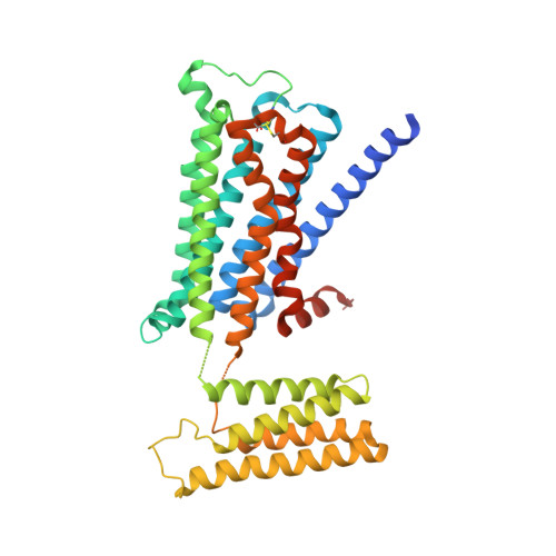
    Human muscarinic receptor M 2 is one of the five subtypes of muscarinic receptors belonging to the family of G-protein-coupled receptors. Muscarinic receptors are targets for multiple neurodegenerative diseases. The challenge has been designing subtype-selective ligands against one of the five muscarinic receptors. We report high-resolution structures of a thermostabilized mutant M 2 receptor bound to a subtype-selective antagonist AF-DX 384 and a nonselective antagonist NMS. The thermostabilizing mutation S110R in M 2 was predicted using a theoretical strategy previously developed in our group. Comparison of the crystal structures and pharmacological properties of the M 2 receptor shows that the Arg in the S110R mutant mimics the stabilizing role of the sodium cation, which is known to allosterically stabilize inactive state(s) of class A GPCRs. Molecular dynamics simulations reveal that tightening of the ligand-residue contacts in M 2 receptors compared to M 3 receptors leads to subtype selectivity of AF-DX 384.


  • Organizational Affiliation

    Department of Cell Biology and Medical Chemistry, Graduate School of Medicine, Kyoto University, Konoe-cho, Yoshida, Sakyo-ku, Kyoto, Japan. r.suno@mfour.med.kyoto-u.ac.jp.


Macromolecules
Find similar proteins by:  (by identity cutoff)  |  3D Structure
Entity ID: 1
MoleculeChains Sequence LengthOrganismDetailsImage
Muscarinic acetylcholine receptor M2,Redesigned apo-cytochrome b562,Muscarinic acetylcholine receptor M2421Homo sapiensMutation(s): 1 
Gene Names: CHRM2
Membrane Entity: Yes 
UniProt & NIH Common Fund Data Resources
Find proteins for P0ABE7 (Escherichia coli)
Explore P0ABE7 
Go to UniProtKB:  P0ABE7
Find proteins for P08172 (Homo sapiens)
Explore P08172 
Go to UniProtKB:  P08172
PHAROS:  P08172
GTEx:  ENSG00000181072 
Entity Groups  
Sequence Clusters30% Identity50% Identity70% Identity90% Identity95% Identity100% Identity
UniProt GroupsP08172P0ABE7
Sequence Annotations
Expand
  • Reference Sequence
Small Molecules
Binding Affinity Annotations 
IDSourceBinding Affinity
3C0 BindingDB:  5YC8 Ki: min: 0.06, max: 10 (nM) from 13 assay(s)
Kd: 0.87 (nM) from 1 assay(s)
IC50: min: 4.3, max: 501 (nM) from 4 assay(s)
Experimental Data & Validation

Experimental Data

  • Method: X-RAY DIFFRACTION
  • Resolution: 2.50 Å
  • R-Value Free:  0.270 (Depositor), 0.270 (DCC) 
  • R-Value Work:  0.237 (Depositor), 0.240 (DCC) 
  • R-Value Observed: 0.239 (Depositor) 
Space Group: P 1 21 1
Unit Cell:
Length ( Å )Angle ( ˚ )
a = 46.52α = 90
b = 59β = 98.89
c = 89.22γ = 90
Software Package:
Software NamePurpose
XSCALEdata scaling
PHENIXrefinement
PDB_EXTRACTdata extraction
XDSdata reduction
PHENIXphasing

Structure Validation

View Full Validation Report


Ligand Structure Quality Assessment 

Created with Raphaël 2.3.0Worse 01 BetterLigand structure goodness of fit to experimental dataBest fitted 3C0Click on this verticalbar to view details

Entry History 

Revision History  (Full details and data files)

  • Version 1.0: 2018-11-21
    Type: Initial release
  • Version 1.1: 2018-11-28
    Changes: Data collection, Database references
  • Version 1.2: 2020-02-26
    Changes: Data collection
  • Version 1.3: 2023-11-22
    Changes: Data collection, Database references, Refinement description
  • Version 1.4: 2024-10-23
    Changes: Structure summary