


Crystal Structure of human TDO inhibitor complex
Fu, G., Wang, J., Luo, G., Wu, G., Qian, K.To be published.
Experimental Data Snapshot
Entity ID: 1 | |||||
|---|---|---|---|---|---|
| Molecule | Chains | Sequence Length | Organism | Details | Image |
| Tryptophan 2,3-dioxygenase | 391 | Homo sapiens | Mutation(s): 0 Gene Names: TDO2, TDO EC: 1.13.11.11 | 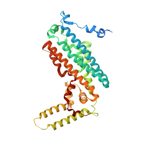 | |
UniProt & NIH Common Fund Data Resources | |||||
PHAROS: P48775 GTEx: ENSG00000151790 | |||||
Entity Groups | |||||
| Sequence Clusters | 30% Identity50% Identity70% Identity90% Identity95% Identity100% Identity | ||||
| UniProt Group | P48775 | ||||
Sequence AnnotationsExpand | |||||
Reference Sequence | |||||
| Ligands 4 Unique | |||||
|---|---|---|---|---|---|
| ID | Chains | Name / Formula / InChI Key | 2D Diagram | 3D Interactions | |
| HEM Download:Ideal Coordinates CCD File | E [auth A], I [auth B], L [auth C], N [auth D] | PROTOPORPHYRIN IX CONTAINING FE C34 H32 Fe N4 O4 KABFMIBPWCXCRK-RGGAHWMASA-L |  | ||
| 9R9 Download:Ideal Coordinates CCD File | F [auth A], J [auth B], M [auth C], O [auth D] | 1-(6-chloro-1H-indazol-4-yl)cyclohexan-1-ol C13 H15 Cl N2 O BMCNEGXEBQNZAQ-UHFFFAOYSA-N |  | ||
| TRP Download:Ideal Coordinates CCD File | G [auth A], K [auth B], P [auth D] | TRYPTOPHAN C11 H12 N2 O2 QIVBCDIJIAJPQS-VIFPVBQESA-N |  | ||
| CIT Download:Ideal Coordinates CCD File | H [auth A] | CITRIC ACID C6 H8 O7 KRKNYBCHXYNGOX-UHFFFAOYSA-N |  | ||
| Length ( Å ) | Angle ( ˚ ) |
|---|---|
| a = 156.308 | α = 90 |
| b = 144.106 | β = 90 |
| c = 89.002 | γ = 90 |
| Software Name | Purpose |
|---|---|
| SCALEPACK | data scaling |
| REFMAC | refinement |
| PDB_EXTRACT | data extraction |
| HKL-2000 | data reduction |
| PHASER | phasing |


