6A9W

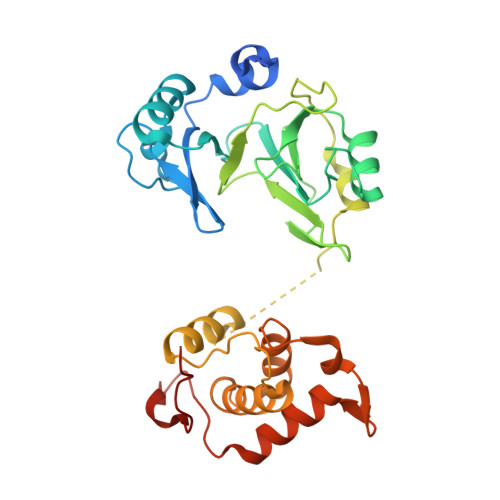
Structure of the bifunctional DNA primase-polymerase from phage NrS-1


Experimental Data Snapshot

  • Method: X-RAY DIFFRACTION
  • Resolution: 1.80 Å
  • R-Value Free: 0.218 
  • R-Value Work: 0.178 
  • R-Value Observed: 0.180 

wwPDB Validation   3D Report Full Report


This is version 1.1 of the entry. See complete history


Literature

Crystal structure and biochemical studies of the bifunctional DNA primase-polymerase from phage NrS-1.

Guo, H.J.Li, M.J.Wang, T.L.Wu, H.Zhou, H.Xu, C.Y.Liu, X.P.Yu, F.He, J.H.

(2019) Biochem Biophys Res Commun 510: 573-579

  • DOI: https://doi.org/10.1016/j.bbrc.2019.01.144
  • Primary Citation of Related Structures:  
    6A9W

  • PubMed Abstract: 

    A novel DNA polymerase found in the deep-sea vent phage NrS-1, was confirmed to have both DNA polymerase and primase activities. In this polymerase, the N-terminal residues 1-300 (referred to as N300) are the core region required for polymerizing DNA and catalyzing de novo DNA synthesis. Here, the crystal structure of N300 was solved at a resolution of 1.80 Å. The overall structure consists of a prim/pol domain and a helix bundle domain, which are separated by a 14-residue-long flexible tether (residues 177-190). Both the prim/pol domain of N300 and other primase-polymerases (prim-pol) encompass an analogous fold with conserved catalytic residues. Mutagenesis and enzymatic activity assays show that the acidic active-site residue E139 is required for both polymerase and primase activities. Functional assays confirm the essentiality of the helix bundle domain for primase activity. Furthermore, we identified a mutant (N300-Y261A) of the helix bundle domain, which probably plays an indispensable role in the primer initiation and recognition of template DNA.


  • Organizational Affiliation

    Shanghai Institute of Applied Physics, Chinese Academy of Sciences, Shanghai, 201800, China; University of Chinese Academy of Sciences, Beijing, 100049, China.


Macromolecules
Find similar proteins by:  (by identity cutoff)  |  3D Structure
Entity ID: 1
MoleculeChains Sequence LengthOrganismDetailsImage
Primase320Nitratiruptor phage NrS-1Mutation(s): 0 
EC: 2.7.7.7 (UniProt), 3.6.4.12 (UniProt), 2.7.7 (UniProt)
UniProt
Find proteins for M5AAG8 (Nitratiruptor phage NrS-1)
Explore M5AAG8 
Go to UniProtKB:  M5AAG8
Entity Groups  
Sequence Clusters30% Identity50% Identity70% Identity90% Identity95% Identity100% Identity
UniProt GroupM5AAG8
Sequence Annotations
Expand
  • Reference Sequence
Experimental Data & Validation

Experimental Data

  • Method: X-RAY DIFFRACTION
  • Resolution: 1.80 Å
  • R-Value Free: 0.218 
  • R-Value Work: 0.178 
  • R-Value Observed: 0.180 
  • Space Group: C 1 2 1
Unit Cell:
Length ( Å )Angle ( ˚ )
a = 119.874α = 90
b = 57.57β = 94.03
c = 47.631γ = 90
Software Package:
Software NamePurpose
PHENIXrefinement
XDSdata reduction
Aimlessdata scaling
PHASERphasing
PDB_EXTRACTdata extraction

Structure Validation

View Full Validation Report


Entry History & Funding Information

Deposition Data


Funding OrganizationLocationGrant Number
National Natural Science Foundation of ChinaChina31570740

Revision History  (Full details and data files)

  • Version 1.0: 2019-03-13
    Type: Initial release
  • Version 1.1: 2024-03-27
    Changes: Data collection, Database references