6DZ5

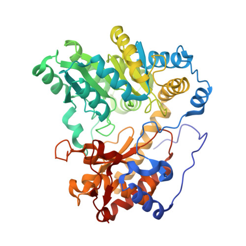
Citrobacter freundii tyrosine phenol-lyase F448A mutant complexed with L-alanine


Experimental Data Snapshot

  • Method: X-RAY DIFFRACTION
  • Resolution: 2.26 Å
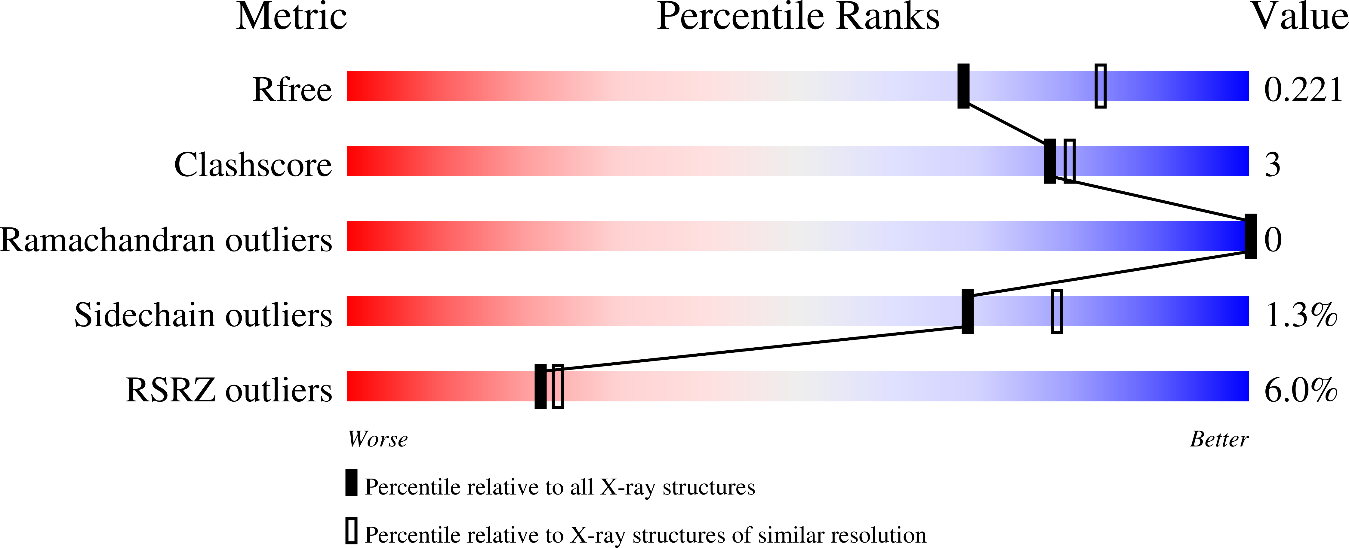
  • R-Value Free: 0.221 
  • R-Value Work: 0.183 
  • R-Value Observed: 0.185 

Starting Model: experimental
View more details

wwPDB Validation   3D Report Full Report


Ligand Structure Quality Assessment 


This is version 1.4 of the entry. See complete history


Literature

Crystal Structures of Wild-Type and F448A Mutant Citrobacter freundii Tyrosine Phenol-Lyase Complexed with a Substrate and Inhibitors: Implications for the Reaction Mechanism.

Phillips, R.S.Craig, S.

(2018) Biochemistry 57: 6166-6179

  • DOI: https://doi.org/10.1021/acs.biochem.8b00724
  • Primary Citation of Related Structures:  
    6DUR, 6DVX, 6DXV, 6DYT, 6DZ5, 6ECG

  • PubMed Abstract: 

    Tyrosine phenol-lyase (TPL; EC 4.1.99.2) is a pyridoxal 5'-phosphate-dependent enzyme that catalyzes the reversible hydrolytic cleavage of l-tyrosine to phenol and ammonium pyruvate. We have shown previously that F448A TPL has k cat and k cat / K m values for l-tyrosine reduced by ∼10 4 -fold [Phillips, R. S., Vita, A., Spivey, J. B., Rudloff, A. P., Driscoll, M. D., and Hay, S. (2016) ACS Catal. 6, 6770-6779]. We have now obtained crystal structures of F448A TPL and complexes with l-alanine, l-methionine, l-phenylalanine, and 3-F-l-tyrosine at 2.05-2.27 Å and the complex of wild-type TPL with l-phenylalanine at 1.8 Å. The small domain of F448A TPL, where Phe-448 is located, is more disordered in chain A than in wild-type TPL. The complexes of F448A TPL with l-alanine and l-phenylalanine are in an open conformation in both chains, while the complex with l-methionine is a 52:48 open:closed equilibrium mixture in chain A. Wild-type TPL with l-alanine is closed in chain A and open in chain B, and the complex with l-phenylalanine is a 56:44 open:closed mixture in chain A. Thus, the Phe-448 to alanine mutation affects the conformational equilibrium of open and closed active sites. The structure of the 3-F-l-tyrosine quinonoid complex of F448A TPL is unstrained and in an open conformation, with a hydrogen bond from the phenolic OH to Thr-124. These results support our previous conclusion that ground-state strain plays a critical role in the mechanism of TPL.


Macromolecules
Find similar proteins by:  (by identity cutoff)  |  3D Structure
Entity ID: 1
MoleculeChains Sequence LengthOrganismDetailsImage
Tyrosine phenol-lyase
A, B
456Citrobacter freundiiMutation(s): 2 
Gene Names: tpl
EC: 4.1.99.2
UniProt
Find proteins for P31013 (Citrobacter freundii)
Explore P31013 
Go to UniProtKB:  P31013
Entity Groups  
Sequence Clusters30% Identity50% Identity70% Identity90% Identity95% Identity100% Identity
UniProt GroupP31013
Sequence Annotations
Expand
  • Reference Sequence
Small Molecules
Ligands 3 Unique
IDChains Name / Formula / InChI Key2D Diagram3D Interactions
P61
Query on P61

Download Ideal Coordinates CCD File 
D [auth A],
G [auth B]
(2E)-3-(3-fluoro-4-hydroxyphenyl)-2-{[(Z)-{3-hydroxy-2-methyl-5-[(phosphonooxy)methyl]pyridin-4(1H)-ylidene}methyl]imino}propanoic acid
C17 H18 F N2 O8 P
AWZHPOMIAYWMIB-ALAGMBGBSA-N
P33
Query on P33

Download Ideal Coordinates CCD File 
F [auth A]3,6,9,12,15,18-HEXAOXAICOSANE-1,20-DIOL
C14 H30 O8
XPJRQAIZZQMSCM-UHFFFAOYSA-N
K
Query on K

Download Ideal Coordinates CCD File 
C [auth A],
E [auth A]
POTASSIUM ION
K
NPYPAHLBTDXSSS-UHFFFAOYSA-N
Experimental Data & Validation

Experimental Data

  • Method: X-RAY DIFFRACTION
  • Resolution: 2.26 Å
  • R-Value Free: 0.221 
  • R-Value Work: 0.183 
  • R-Value Observed: 0.185 
  • Space Group: P 2 21 21
Unit Cell:
Length ( Å )Angle ( ˚ )
a = 59.88α = 90
b = 132.81β = 90
c = 145.37γ = 90
Software Package:
Software NamePurpose
PHENIXrefinement
XSCALEdata scaling
PDB_EXTRACTdata extraction
XDSdata reduction
PHASERphasing

Structure Validation

View Full Validation Report


Ligand Structure Quality Assessment 


Entry History 

Deposition Data

Revision History  (Full details and data files)

  • Version 1.0: 2018-10-10
    Type: Initial release
  • Version 1.1: 2018-11-14
    Changes: Data collection, Database references, Structure summary
  • Version 1.2: 2019-03-06
    Changes: Data collection, Database references, Structure summary
  • Version 1.3: 2019-10-23
    Changes: Data collection, Database references
  • Version 1.4: 2023-10-11
    Changes: Data collection, Database references, Derived calculations, Refinement description, Structure summary