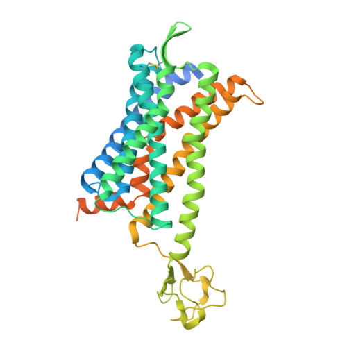
6GPS

CRYSTAL STRUCTURE OF CCR2A IN COMPLEX WITH MK-0812


Experimental Data Snapshot

  • Method: X-RAY DIFFRACTION
  • Resolution: 3.30 Å
  • R-Value Free: 0.296 
  • R-Value Work: 0.243 
  • R-Value Observed: 0.245 

wwPDB Validation   3D Report Full Report


Ligand Structure Quality Assessment 


This is version 1.2 of the entry. See complete history


Literature

Crystal Structure of CC Chemokine Receptor 2A in Complex with an Orthosteric Antagonist Provides Insights for the Design of Selective Antagonists.

Apel, A.K.Cheng, R.K.Y.Tautermann, C.S.Brauchle, M.Huang, C.Y.Pautsch, A.Hennig, M.Nar, H.Schnapp, G.

(2019) Structure 27: 427-438.e5

  • DOI: https://doi.org/10.1016/j.str.2018.10.027
  • Primary Citation of Related Structures:  
    6GPS, 6GPX

  • PubMed Abstract: 

    We determined two crystal structures of the chemokine receptor CCR2A in complex with the orthosteric antagonist MK-0812. Full-length CCR2A, stabilized by rubredoxin and a series of five mutations were resolved at 3.3 Å. An N- and C-terminally truncated CCR2A construct was crystallized in an alternate crystal form, which yielded a 2.7 Å resolution structure using serial synchrotron crystallography. Our structures provide a clear structural explanation for the observed key role of residue E291 7.39 in high-affinity binding of several orthosteric CCR2 antagonists. By combining all the structural information collected, we generated models of co-structures for the structurally diverse pyrimidine amide class of CCR2 antagonists. Even though the representative Ex15 overlays well with MK-0812, it also interacts with the non-conserved H121 3.33 , resulting in a significant selectivity over CCR5. Insights derived from this work will facilitate drug discovery efforts directed toward highly selective CCR2 antagonists with potentially superior efficacy.


  • Organizational Affiliation

    Boehringer Ingelheim Pharma GmbH & Co. KG, Birkendorferstrasse 65, 88397 Biberach, Germany.


Macromolecules
Find similar proteins by:  (by identity cutoff)  |  3D Structure
Entity ID: 1
MoleculeChains Sequence LengthOrganismDetailsImage
C-C chemokine receptor type 2,Rubredoxin,C-C chemokine receptor type 2429Homo sapiensClostridium pasteurianumMutation(s): 5 
Gene Names: CCR2CMKBR2
Membrane Entity: Yes 
UniProt & NIH Common Fund Data Resources
Find proteins for P41597 (Homo sapiens)
Explore P41597 
Go to UniProtKB:  P41597
PHAROS:  P41597
GTEx:  ENSG00000121807 
Find proteins for P00268 (Clostridium pasteurianum)
Explore P00268 
Go to UniProtKB:  P00268
Entity Groups  
Sequence Clusters30% Identity50% Identity70% Identity90% Identity95% Identity100% Identity
UniProt GroupsP00268P41597
Sequence Annotations
Expand
  • Reference Sequence
Small Molecules
Ligands 2 Unique
IDChains Name / Formula / InChI Key2D Diagram3D Interactions
F7N
Query on F7N

Download Ideal Coordinates CCD File 
C [auth A][(3~{S},4~{S})-3-methoxyoxan-4-yl]-[(1~{R},3~{S})-3-propan-2-yl-3-[[3-(trifluoromethyl)-7,8-dihydro-5~{H}-1,6-naphthyridin-6-yl]carbonyl]cyclopentyl]azanium
C24 H35 F3 N3 O3
MTMDXAIUENDNDL-RJSMDTJLSA-O
ZN
Query on ZN

Download Ideal Coordinates CCD File 
B [auth A]ZINC ION
Zn
PTFCDOFLOPIGGS-UHFFFAOYSA-N
Experimental Data & Validation

Experimental Data

  • Method: X-RAY DIFFRACTION
  • Resolution: 3.30 Å
  • R-Value Free: 0.296 
  • R-Value Work: 0.243 
  • R-Value Observed: 0.245 
  • Space Group: P 1 21 1
Unit Cell:
Length ( Å )Angle ( ˚ )
a = 38.5α = 90
b = 61.2β = 97.66
c = 123.57γ = 90
Software Package:
Software NamePurpose
BUSTERrefinement
XDSdata reduction
autoPROCdata scaling
PHASERphasing

Structure Validation

View Full Validation Report


Ligand Structure Quality Assessment 


Entry History 

Deposition Data

Revision History  (Full details and data files)

  • Version 1.0: 2019-01-02
    Type: Initial release
  • Version 1.1: 2019-03-13
    Changes: Data collection, Database references
  • Version 1.2: 2024-11-20
    Changes: Data collection, Database references, Refinement description, Structure summary