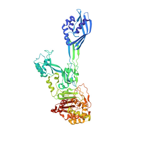
6H5O

Crystal structure of PBP2a from MRSA in complex with piperacillin at active site.


Experimental Data Snapshot

  • Method: X-RAY DIFFRACTION
  • Resolution: 2.82 Å
  • R-Value Free: 0.311 
  • R-Value Work: 0.241 
  • R-Value Observed: 0.244 

Starting Model: experimental
View more details

wwPDB Validation   3D Report Full Report


Ligand Structure Quality Assessment 


This is version 1.4 of the entry. See complete history


Literature

The Quinazolinone Allosteric Inhibitor of PBP 2a Synergizes with Piperacillin and Tazobactam against Methicillin-Resistant Staphylococcus aureus.

Janardhanan, J.Bouley, R.Martinez-Caballero, S.Peng, Z.Batuecas-Mordillo, M.Meisel, J.E.Ding, D.Schroeder, V.A.Wolter, W.R.Mahasenan, K.V.Hermoso, J.A.Mobashery, S.Chang, M.

(2019) Antimicrob Agents Chemother 63

  • DOI: https://doi.org/10.1128/AAC.02637-18
  • Primary Citation of Related Structures:  
    6H5O, 6Q9N

  • PubMed Abstract: 

    The quinazolinones are a new class of antibacterials with in vivo efficacy against methicillin-resistant Staphylococcus aureus (MRSA). The quinazolinones target cell wall biosynthesis and have a unique mechanism of action by binding to the allosteric site of penicillin-binding protein 2a (PBP 2a). We investigated the potential for synergism of a lead quinazolinone with several antibiotics of different classes using checkerboard and time-kill assays. The quinazolinone synergized with β-lactam antibiotics. The combination of the quinazolinone with commercial piperacillin-tazobactam showed bactericidal synergy at sub-MICs of all three drugs. We demonstrated the efficacy of the triple-drug combination in a mouse MRSA neutropenic thigh infection model. The proposed mechanism for the synergistic activity in MRSA involves inhibition of the β-lactamase by tazobactam, which protects piperacillin from hydrolysis, which can then inhibit its target, PBP 2. Furthermore, the quinazolinone binds to the allosteric site of PBP 2a, triggering the allosteric response. This leads to the opening of the active site, which, in turn, binds another molecule of piperacillin. In other words, PBP 2a, which is not normally inhibited by piperacillin, becomes vulnerable to inhibition in the presence of the quinazolinone. The collective effect is the impairment of cell wall biosynthesis, with bactericidal consequence. Two crystal structures for complexes of the antibiotics with PBP 2a provide support for the proposed mechanism of action.


  • Organizational Affiliation

    Department of Chemistry and Biochemistry, University of Notre Dame, Notre Dame, Indiana, USA.


Macromolecules
Find similar proteins by:  (by identity cutoff)  |  3D Structure
Entity ID: 1
MoleculeChains Sequence LengthOrganismDetailsImage
Penicillin binding protein 2 prime
A, B
642Staphylococcus aureus subsp. aureus Mu50Mutation(s): 0 
EC: 3.4.16.4
UniProt
Find proteins for A0A0H3JPA5 (Staphylococcus aureus (strain Mu50 / ATCC 700699))
Explore A0A0H3JPA5 
Go to UniProtKB:  A0A0H3JPA5
Entity Groups  
Sequence Clusters30% Identity50% Identity70% Identity90% Identity95% Identity100% Identity
UniProt GroupA0A0H3JPA5
Sequence Annotations
Expand
  • Reference Sequence
Small Molecules
Ligands 2 Unique
IDChains Name / Formula / InChI Key2D Diagram3D Interactions
JPP
Query on JPP

Download Ideal Coordinates CCD File 
C [auth A],
L [auth B]
Piperacillin (Open Form)
C23 H29 N5 O7 S
CJYMHKMECTVNSA-XLMAVXFVSA-N
CD
Query on CD

Download Ideal Coordinates CCD File 
D [auth A]
E [auth A]
F [auth A]
G [auth A]
H [auth A]
D [auth A],
E [auth A],
F [auth A],
G [auth A],
H [auth A],
I [auth A],
J [auth A],
K [auth A],
M [auth B]
CADMIUM ION
Cd
WLZRMCYVCSSEQC-UHFFFAOYSA-N
Experimental Data & Validation

Experimental Data

  • Method: X-RAY DIFFRACTION
  • Resolution: 2.82 Å
  • R-Value Free: 0.311 
  • R-Value Work: 0.241 
  • R-Value Observed: 0.244 
  • Space Group: P 21 21 21
Unit Cell:
Length ( Å )Angle ( ˚ )
a = 80.713α = 90
b = 105.014β = 90
c = 185.624γ = 90
Software Package:
Software NamePurpose
REFMACrefinement
XDSdata reduction
Aimlessdata scaling
PHASERphasing

Structure Validation

View Full Validation Report


Ligand Structure Quality Assessment 


Entry History & Funding Information

Deposition Data


Funding OrganizationLocationGrant Number
National Institutes of Health/National Institute Of Allergy and Infectious Diseases (NIH/NIAID)United States1R01AI116548

Revision History  (Full details and data files)

  • Version 1.0: 2019-08-14
    Type: Initial release
  • Version 1.1: 2020-09-09
    Changes: Database references, Derived calculations
  • Version 1.2: 2022-03-30
    Changes: Author supporting evidence, Database references
  • Version 1.3: 2024-01-17
    Changes: Data collection, Refinement description
  • Version 1.4: 2024-10-16
    Changes: Structure summary