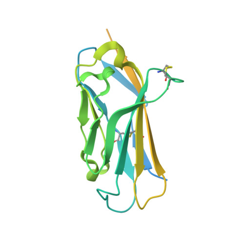
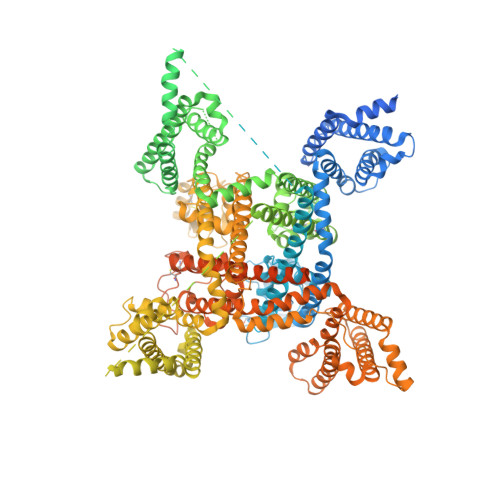
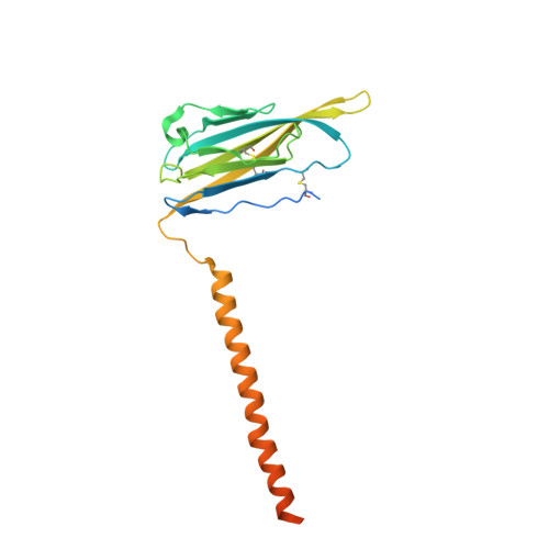
6J8G

Structure of human voltage-gated sodium channel Nav1.7 in complex with auxiliary beta subunits, huwentoxin-IV and saxitoxin (Y1755 up)


Experimental Data Snapshot

  • Method: ELECTRON MICROSCOPY
  • Resolution: 3.20 Å
  • Aggregation State: PARTICLE 
  • Reconstruction Method: SINGLE PARTICLE 

wwPDB Validation   3D Report Full Report


This is version 2.1 of the entry. See complete history


Literature

Structures of human Nav1.7 channel in complex with auxiliary subunits and animal toxins.

Shen, H.Liu, D.Wu, K.Lei, J.Yan, N.

(2019) Science 363: 1303-1308

  • DOI: https://doi.org/10.1126/science.aaw2493
  • Primary Citation of Related Structures:  
    6J8G, 6J8H, 6J8I, 6J8J

  • PubMed Abstract: 

    Voltage-gated sodium channel Na v 1.7 represents a promising target for pain relief. Here we report the cryo-electron microscopy structures of the human Na v 1.7-β1-β2 complex bound to two combinations of pore blockers and gating modifier toxins (GMTs), tetrodotoxin with protoxin-II and saxitoxin with huwentoxin-IV, both determined at overall resolutions of 3.2 angstroms. The two structures are nearly identical except for minor shifts of voltage-sensing domain II (VSD II ), whose S3-S4 linker accommodates the two GMTs in a similar manner. One additional protoxin-II sits on top of the S3-S4 linker in VSD IV The structures may represent an inactivated state with all four VSDs "up" and the intracellular gate closed. The structures illuminate the path toward mechanistic understanding of the function and disease of Na v 1.7 and establish the foundation for structure-aided development of analgesics.


  • Organizational Affiliation

    State Key Laboratory of Membrane Biology, Tsinghua University, Beijing 100084, China.


Macromolecules
Find similar proteins by:  (by identity cutoff)  |  3D Structure
Entity ID: 1
MoleculeChains Sequence LengthOrganismDetailsImage
Sodium channel subunit beta-2A [auth C]215Homo sapiensMutation(s): 0 
Gene Names: SCN2BUNQ326/PRO386
Membrane Entity: Yes 
UniProt & NIH Common Fund Data Resources
Find proteins for O60939 (Homo sapiens)
Explore O60939 
Go to UniProtKB:  O60939
PHAROS:  O60939
GTEx:  ENSG00000149575 
Entity Groups  
Sequence Clusters30% Identity50% Identity70% Identity90% Identity95% Identity100% Identity
UniProt GroupO60939
Glycosylation
Glycosylation Sites: 1Go to GlyGen: O60939-1
Sequence Annotations
Expand
  • Reference Sequence
Find similar proteins by:  (by identity cutoff)  |  3D Structure
Entity ID: 2
MoleculeChains Sequence LengthOrganismDetailsImage
Sodium channel protein type 9 subunit alphaB [auth A]2,031Homo sapiensMutation(s): 1 
Gene Names: SCN9ANENA
Membrane Entity: Yes 
UniProt & NIH Common Fund Data Resources
Find proteins for Q15858 (Homo sapiens)
Explore Q15858 
Go to UniProtKB:  Q15858
PHAROS:  Q15858
GTEx:  ENSG00000169432 
Entity Groups  
Sequence Clusters30% Identity50% Identity70% Identity90% Identity95% Identity100% Identity
UniProt GroupQ15858
Glycosylation
Glycosylation Sites: 4Go to GlyGen: Q15858-1
Sequence Annotations
Expand
  • Reference Sequence
Find similar proteins by:  (by identity cutoff)  |  3D Structure
Entity ID: 3
MoleculeChains Sequence LengthOrganismDetailsImage
Sodium channel subunit beta-1C [auth B]218Homo sapiensMutation(s): 0 
Gene Names: SCN1B
Membrane Entity: Yes 
UniProt & NIH Common Fund Data Resources
Find proteins for Q07699 (Homo sapiens)
Explore Q07699 
Go to UniProtKB:  Q07699
PHAROS:  Q07699
GTEx:  ENSG00000105711 
Entity Groups  
Sequence Clusters30% Identity50% Identity70% Identity90% Identity95% Identity100% Identity
UniProt GroupQ07699
Glycosylation
Glycosylation Sites: 4Go to GlyGen: Q07699-1
Sequence Annotations
Expand
  • Reference Sequence
Oligosaccharides

Help

Entity ID: 4
MoleculeChains Length2D Diagram Glycosylation3D Interactions
2-acetamido-2-deoxy-beta-D-glucopyranose-(1-4)-2-acetamido-2-deoxy-beta-D-glucopyranose
D, E
2N-Glycosylation
Glycosylation Resources
GlyTouCan:  G42666HT
GlyCosmos:  G42666HT
GlyGen:  G42666HT
Small Molecules
Ligands 2 Unique
IDChains Name / Formula / InChI Key2D Diagram3D Interactions
9SL
Query on 9SL

Download Ideal Coordinates CCD File 
I [auth A][(3aS,4R,10aS)-2,6-diamino-10,10-dihydroxy-3a,4,9,10-tetrahydro-3H,8H-pyrrolo[1,2-c]purin-4-yl]methyl carbamate
C10 H17 N7 O4
RPQXVSUAYFXFJA-HGRQIUPRSA-N
NAG
Query on NAG

Download Ideal Coordinates CCD File 
F [auth C]
G [auth A]
H [auth A]
J [auth B]
K [auth B]
F [auth C],
G [auth A],
H [auth A],
J [auth B],
K [auth B],
L [auth B],
M [auth B]
2-acetamido-2-deoxy-beta-D-glucopyranose
C8 H15 N O6
OVRNDRQMDRJTHS-FMDGEEDCSA-N
Experimental Data & Validation

Experimental Data

  • Method: ELECTRON MICROSCOPY
  • Resolution: 3.20 Å
  • Aggregation State: PARTICLE 
  • Reconstruction Method: SINGLE PARTICLE 

Structure Validation

View Full Validation Report


Entry History & Funding Information

Deposition Data


Funding OrganizationLocationGrant Number
Ministry of Science and Technology (China)China2015CB910101
Ministry of Science and Technology (China)China2016YFA0500402
National Natural Science Foundation of ChinaChina31621092
National Natural Science Foundation of ChinaChina31630017
National Natural Science Foundation of ChinaChina81861138009

Revision History  (Full details and data files)

  • Version 1.0: 2019-02-27
    Type: Initial release
  • Version 1.1: 2019-04-10
    Changes: Data collection, Database references
  • Version 1.2: 2019-11-06
    Changes: Data collection, Other
  • Version 2.0: 2020-07-29
    Type: Remediation
    Reason: Carbohydrate remediation
    Changes: Advisory, Atomic model, Data collection, Derived calculations, Structure summary
  • Version 2.1: 2024-11-06
    Changes: Data collection, Database references, Structure summary