6JMQ | pdb_00006jmq

LAT1-CD98hc complex bound to MEM-108 Fab


Experimental Data Snapshot

  • Method: ELECTRON MICROSCOPY
  • Resolution: 3.31 Å
  • Aggregation State: PARTICLE 
  • Reconstruction Method: SINGLE PARTICLE 

wwPDB Validation 3D Report Full Report

Validation slider image for 6JMQ

This is version 2.2 of the entry. See complete history

Literature

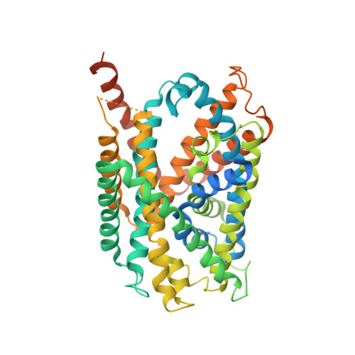
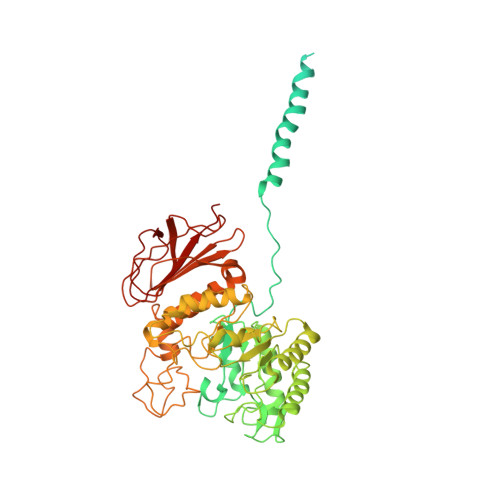
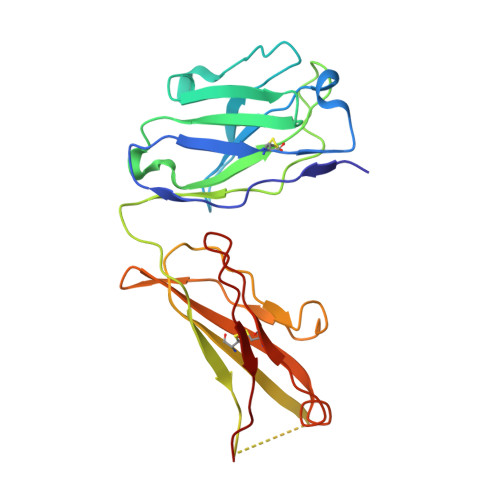
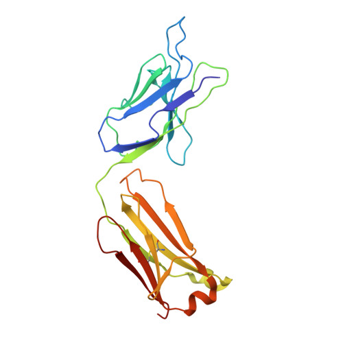
Cryo-EM structure of the human L-type amino acid transporter 1 in complex with glycoprotein CD98hc.

Lee, Y.Wiriyasermkul, P.Jin, C.Quan, L.Ohgaki, R.Okuda, S.Kusakizako, T.Nishizawa, T.Oda, K.Ishitani, R.Yokoyama, T.Nakane, T.Shirouzu, M.Endou, H.Nagamori, S.Kanai, Y.Nureki, O.

(2019) Nat Struct Mol Biol 26: 510-517

  • DOI: https://doi.org/10.1038/s41594-019-0237-7
  • Primary Citation Related Structures: 
    6JMQ, 6JMR

  • PubMed Abstract: 

    The L-type amino acid transporter 1 (LAT1 or SLC7A5) transports large neutral amino acids across the membrane and is crucial for brain drug delivery and tumor growth. LAT1 forms a disulfide-linked heterodimer with CD98 heavy chain (CD98hc, 4F2hc or SLC3A2), but the mechanism of assembly and amino acid transport are poorly understood. Here we report the cryo-EM structure of the human LAT1-CD98hc heterodimer at 3.3-Å resolution. LAT1 features a canonical Leu T-fold and exhibits an unusual loop structure on transmembrane helix 6, creating an extended cavity that might accommodate bulky amino acids and drugs. CD98hc engages with LAT1 through the extracellular, transmembrane and putative cholesterol-mediated interactions. We also show that two anti-CD98 antibodies recognize distinct, multiple epitopes on CD98hc but not its glycans, explaining their robust reactivities. These results reveal the principles of glycoprotein-solute carrier assembly and provide templates for improving preclinical drugs and antibodies targeting LAT1 or CD98hc.


  • Organizational Affiliation
    • Department of Biological Sciences, Graduate School of Science, The University of Tokyo, Tokyo, Japan.

Macromolecule Content 

  • Total Structure Weight: 172.94 kDa 
  • Atom Count: 9,950 
  • Modeled Residue Count: 1,287 
  • Deposited Residue Count: 1,584 
  • Unique protein chains: 4

Macromolecules

Find similar proteins by:|  3D Structure
Entity ID: 1
MoleculeChains  Sequence LengthOrganismDetailsImage
Large neutral amino acids transporter small subunit 1515Homo sapiensMutation(s): 0 
Gene Names: SLC7A5CD98LCLAT1MPE16
Membrane Entity: Yes 
UniProt & NIH Common Fund Data Resources
Find proteins for Q01650 (Homo sapiens)
Explore Q01650 
Go to UniProtKB:  Q01650
PHAROS:  Q01650
GTEx:  ENSG00000103257 
Entity Groups
Sequence Clusters30% Identity50% Identity70% Identity90% Identity95% Identity100% Identity
UniProt GroupQ01650
Sequence Annotations
Expand
Reference Sequence
Find similar proteins by:|  3D Structure
Entity ID: 2
MoleculeChains  Sequence LengthOrganismDetailsImage
4F2 cell-surface antigen heavy chain631Homo sapiensMutation(s): 0 
Gene Names: SLC3A2MDU1
Membrane Entity: Yes 
UniProt & NIH Common Fund Data Resources
Find proteins for P08195 (Homo sapiens)
Explore P08195 
Go to UniProtKB:  P08195
PHAROS:  P08195
GTEx:  ENSG00000168003 
Entity Groups
Sequence Clusters30% Identity50% Identity70% Identity90% Identity95% Identity100% Identity
UniProt GroupP08195
Glycosylation
Glycosylation Sites: 4Go to GlyGen: P08195-1
Sequence Annotations
Expand
Reference Sequence
Find similar proteins by:|  3D Structure
Entity ID: 3
MoleculeChains  Sequence LengthOrganismDetailsImage
Antibody219Mus musculusMutation(s): 0 
Find similar proteins by:|  3D Structure
Entity ID: 4
MoleculeChains  Sequence LengthOrganismDetailsImage
Antibody219Mus musculusMutation(s): 0 

Oligosaccharides

Help  
Entity ID: 5
MoleculeChains Length2D Diagram GlycosylationD Interactions
2-acetamido-2-deoxy-beta-D-glucopyranose-(1-4)-2-acetamido-2-deoxy-beta-D-glucopyranose
E
2N-Glycosylation
Glycosylation Resources
GlyTouCan: G42666HT
GlyCosmos: G42666HT
GlyGen: G42666HT

Small Molecules

Ligands 2 Unique
IDChains Name / Formula / InChI Key2D Diagram3D Interactions
CLR

Query on CLR


Download:Ideal Coordinates CCD File
F [auth A],
G [auth A],
H [auth A],
I [auth A],
J [auth B]
CHOLESTEROL
C27 H46 O
HVYWMOMLDIMFJA-DPAQBDIFSA-N
NAG

Query on NAG


Download:Ideal Coordinates CCD File
K [auth B],
L [auth B],
M [auth B]
2-acetamido-2-deoxy-beta-D-glucopyranose
C8 H15 N O6
OVRNDRQMDRJTHS-FMDGEEDCSA-N

Experimental Data & Validation

Experimental Data

  • Method: ELECTRON MICROSCOPY
  • Resolution: 3.31 Å
  • Aggregation State: PARTICLE 
  • Reconstruction Method: SINGLE PARTICLE 
EM Software:
TaskSoftware PackageVersion
MODEL REFINEMENTPHENIX

Structure Validation

View Full Validation Report


Entry History 

& Funding Information

Deposition Data


Funding OrganizationLocationGrant Number
Japan Society for the Promotion of ScienceJapanJP16J07405
Japan Agency for Medical Research and Development (AMED)JapanJP18am0101115

Revision History  (Full details and data files)

  • Version 1.0: 2019-06-19
    Type: Initial release
  • Version 1.1: 2019-11-06
    Changes: Data collection, Other
  • Version 2.0: 2020-07-29
    Type: Remediation
    Reason: Carbohydrate remediation
    Changes: Atomic model, Data collection, Derived calculations, Structure summary
  • Version 2.1: 2024-10-30
    Changes: Data collection, Database references, Structure summary
  • Version 2.2: 2025-06-25
    Changes: Data collection