6KU3 | pdb_00006ku3

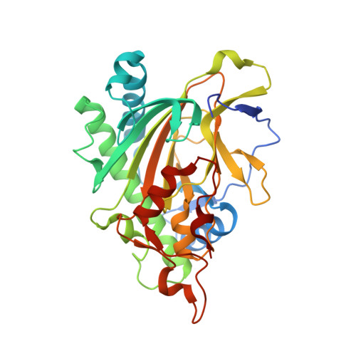
Crystal structure of gibberellin 2-oxidase3 (GA2ox3)in rice


Experimental Data Snapshot

  • Method: X-RAY DIFFRACTION
  • Resolution: 2.15 Å
  • R-Value Free: 
    0.243 (Depositor), 0.246 (DCC) 
  • R-Value Work: 
    0.197 (Depositor), 0.208 (DCC) 
  • R-Value Observed: 
    0.199 (Depositor) 

wwPDB Validation 3D Report Full Report

Validation slider image for 6KU3

Ligand Structure Quality Assessment 


This is version 1.1 of the entry. See complete history. 

Literature

A common allosteric mechanism regulates homeostatic inactivation of auxin and gibberellin.

Takehara, S., Sakuraba, S., Mikami, B., Yoshida, H., Yoshimura, H., Itoh, A., Endo, M., Watanabe, N., Nagae, T., Matsuoka, M., Ueguchi-Tanaka, M.

(2020) Nat Commun 11: 2143-2143

  • DOI: https://doi.org/10.1038/s41467-020-16068-0
  • Primary Citation Related Structures: 
    6KU3, 6KUN

  • PubMed Abstract: 

    Allosteric regulation is protein activation by effector binding at a site other than the active site. Here, we show via X-ray structural analysis of gibberellin 2-oxidase 3 (GA2ox3), and auxin dioxygenase (DAO), that such a mechanism maintains hormonal homeostasis in plants. Both enzymes form multimers by interacting via GA 4 and indole-3-acetic acid (IAA) at their binding interface. Via further functional analyses we reveal that multimerization of these enzymes gradually proceeds with increasing GA 4 and IAA concentrations; multimerized enzymes have higher specific activities than monomer forms, a system that should favour the maintenance of homeostasis for these phytohormones. Molecular dynamic analysis suggests a possible mechanism underlying increased GA2ox3 activity by multimerization-GA 4 in the interface of oligomerized GA2ox3s may be able to enter the active site with a low energy barrier. In summary, homeostatic systems for maintaining GA and IAA levels, based on a common allosteric mechanism, appear to have developed independently.


  • Organizational Affiliation: 
    • Bioscience and Biotechnology Centre, Nagoya University, Nagoya, 464-8601, Japan.

Macromolecule Content 

  • Total Structure Weight: 145.54 kDa 
  • Atom Count: 10,323 
  • Modeled Residue Count: 1,262 
  • Deposited Residue Count: 1,308 
  • Unique protein chains: 1

Macromolecules

Find similar proteins by:|  3D Structure
Entity ID: 1
MoleculeChains  Sequence LengthOrganismDetailsImage
Gibberellin 2-beta-dioxygenase 3
A, B, C, D
327Oryza sativa Japonica GroupMutation(s): 0 
Gene Names: GA2OX3, Os01g0757200, LOC_Os01g55240, OJ1414_E05.17
EC: 1.14.11.13
UniProt
Find proteins for Q8S0S6 (Oryza sativa subsp. japonica)
Explore Q8S0S6 
Go to UniProtKB:  Q8S0S6
Entity Groups
Sequence Clusters30% Identity50% Identity70% Identity90% Identity95% Identity100% Identity
UniProt GroupQ8S0S6
Sequence Annotations
Expand
Reference Sequence

Small Molecules

Ligands 4 Unique
IDChains Name / Formula / InChI Key2D Diagram3D Interactions
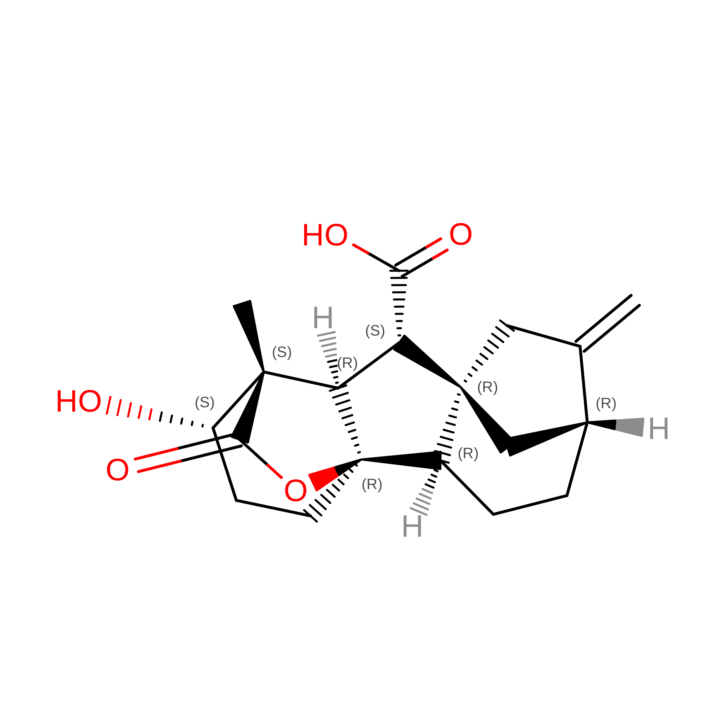
GA4
(Subject of Investigation/LOI)

Query on GA4


Download:Ideal Coordinates CCD File
E [auth A]
F [auth A]
O [auth B]
U [auth C]
V [auth C]
E [auth A],
F [auth A],
O [auth B],
U [auth C],
V [auth C],
Z [auth D]
GIBBERELLIN A4
C19 H24 O5
RSQSQJNRHICNNH-NFMPGMCNSA-N
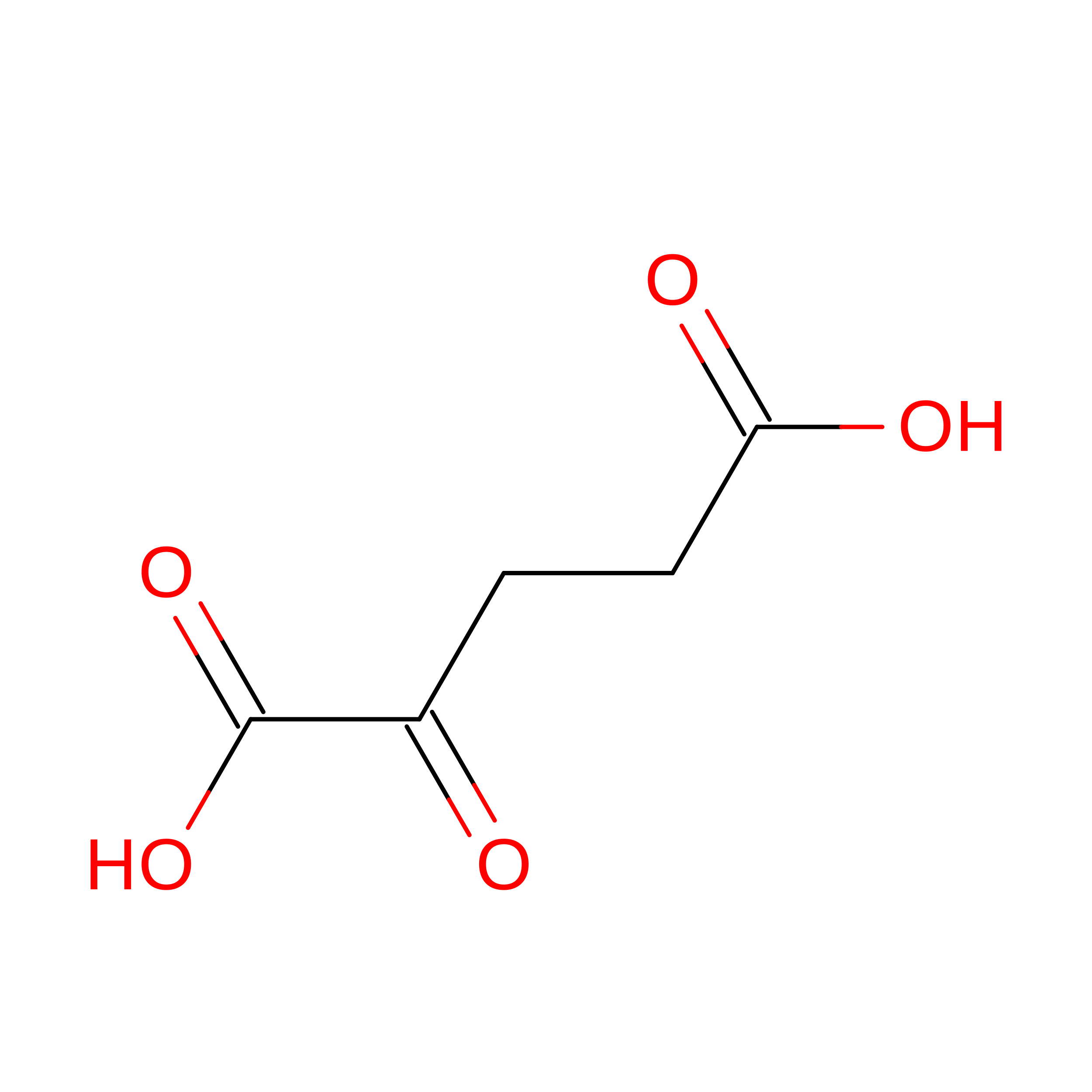
AKG

Query on AKG


Download:Ideal Coordinates CCD File
AA [auth D],
G [auth A],
P [auth B],
W [auth C]
2-OXOGLUTARIC ACID
C5 H6 O5
KPGXRSRHYNQIFN-UHFFFAOYSA-N
SO4

Query on SO4


Download:Ideal Coordinates CCD File
DA [auth D]
L [auth A]
M [auth A]
N [auth A]
T [auth B]
DA [auth D],
L [auth A],
M [auth A],
N [auth A],
T [auth B],
Y [auth C]
SULFATE ION
O4 S
QAOWNCQODCNURD-UHFFFAOYSA-L
GOL

Query on GOL


Download:Ideal Coordinates CCD File
BA [auth D]
CA [auth D]
H [auth A]
I [auth A]
J [auth A]
BA [auth D],
CA [auth D],
H [auth A],
I [auth A],
J [auth A],
K [auth A],
Q [auth B],
R [auth B],
S [auth B],
X [auth C]
GLYCEROL
C3 H8 O3
PEDCQBHIVMGVHV-UHFFFAOYSA-N

Experimental Data & Validation

Experimental Data

  • Method: X-RAY DIFFRACTION
  • Resolution: 2.15 Å
  • R-Value Free:  0.243 (Depositor), 0.246 (DCC) 
  • R-Value Work:  0.197 (Depositor), 0.208 (DCC) 
  • R-Value Observed: 0.199 (Depositor) 
Space Group: P 21 21 21
Unit Cell:
Length ( Å )Angle ( ˚ )
a = 99.47α = 90
b = 112.739β = 90
c = 149.51γ = 90
Software Package:
Software NamePurpose
PHENIXrefinement
Cootmodel building
HKL-2000data reduction
HKL-2000data scaling
HKL-2000data collection
MOLREPphasing

Structure Validation

View Full Validation Report


Ligand Structure Quality Assessment 


Entry History 

& Funding Information

Deposition Data


Funding OrganizationLocationGrant Number
Japan Society for the Promotion of ScienceJapan16H06464
Japan Society for the Promotion of ScienceJapan16H06468

Revision History  (Full details and data files)

  • Version 1.0: 2020-05-13
    Type: Initial release
  • Version 1.1: 2024-10-23
    Changes: Data collection, Database references, Structure summary