


Computationally-guided design and affinity improvement of a protein binder targeting a specific site on HER2
Kim, T.Y., Cha, J.S., Kim, H., Choi, Y., Cho, H.S., Kim, H.S.(2021) Comput Struct Biotechnol J 19: 1325-1334
Experimental Data Snapshot
Starting Model: experimental
View more details
Entity ID: 1 | |||||
|---|---|---|---|---|---|
| Molecule | Chains | Sequence Length | Organism | Details | Image |
| Repebody (Rb-H2) | 275 | Cyclostomata | Mutation(s): 0 | 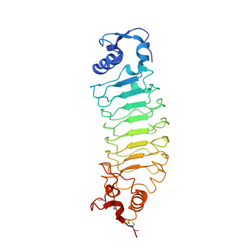 | |
Entity ID: 2 | |||||
|---|---|---|---|---|---|
| Molecule | Chains | Sequence Length | Organism | Details | Image |
| Receptor tyrosine-protein kinase erbB-2 | 97 | Homo sapiens | Mutation(s): 0 Gene Names: ERBB2, HER2, MLN19, NEU, NGL EC: 2.7.10.1 | 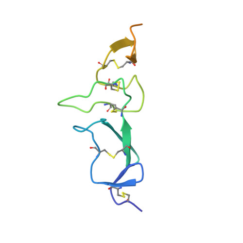 | |
UniProt & NIH Common Fund Data Resources | |||||
PHAROS: P04626 GTEx: ENSG00000141736 | |||||
Entity Groups | |||||
| Sequence Clusters | 30% Identity50% Identity70% Identity90% Identity95% Identity100% Identity | ||||
| UniProt Group | P04626 | ||||
Glycosylation | |||||
| Glycosylation Sites: 1 | Go to GlyGen: P04626-1 | ||||
Sequence AnnotationsExpand | |||||
Reference Sequence | |||||
| Ligands 1 Unique | |||||
|---|---|---|---|---|---|
| ID | Chains | Name / Formula / InChI Key | 2D Diagram | 3D Interactions | |
| NAG Download:Ideal Coordinates CCD File | C [auth B] | 2-acetamido-2-deoxy-beta-D-glucopyranose C8 H15 N O6 OVRNDRQMDRJTHS-FMDGEEDCSA-N |  | ||
| Length ( Å ) | Angle ( ˚ ) |
|---|---|
| a = 44.659 | α = 90 |
| b = 80.071 | β = 90 |
| c = 108.406 | γ = 90 |
| Software Name | Purpose |
|---|---|
| PHENIX | refinement |
| XDS | data reduction |
| XDS | data scaling |
| PHASER | phasing |


