


Structure-based Optimization of Potent, Cell-Active Hydroxylactam Inhibitors of Lactate Dehydrogenase
Wei, B., Robarge, K., Labadie, S.S., Chen, J., Corson, L.B., DiPasquale, A., Eigenbrot, C., Ultsch, M.To be published.
Experimental Data Snapshot
Entity ID: 1 | |||||
|---|---|---|---|---|---|
| Molecule | Chains | Sequence Length | Organism | Details | Image |
| L-lactate dehydrogenase A chain | 332 | Homo sapiens | Mutation(s): 0 Gene Names: LDHA, PIG19 EC: 1.1.1.27 | 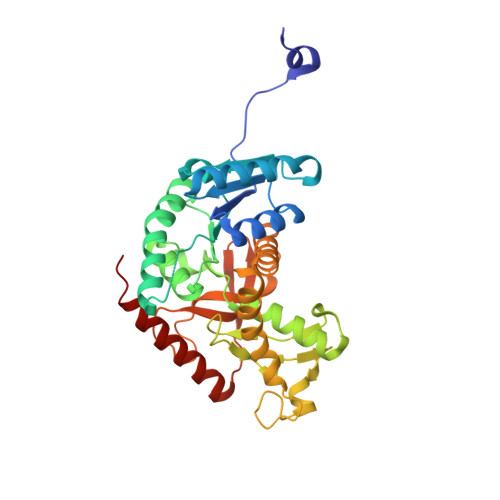 | |
UniProt & NIH Common Fund Data Resources | |||||
PHAROS: P00338 GTEx: ENSG00000134333 | |||||
Entity Groups | |||||
| Sequence Clusters | 30% Identity50% Identity70% Identity90% Identity95% Identity100% Identity | ||||
| UniProt Group | P00338 | ||||
Sequence AnnotationsExpand | |||||
Reference Sequence | |||||
| Ligands 4 Unique | |||||
|---|---|---|---|---|---|
| ID | Chains | Name / Formula / InChI Key | 2D Diagram | 3D Interactions | |
| NAI Download:Ideal Coordinates CCD File | E [auth A], I [auth B], L [auth C], O [auth D] | 1,4-DIHYDRONICOTINAMIDE ADENINE DINUCLEOTIDE C21 H29 N7 O14 P2 BOPGDPNILDQYTO-NNYOXOHSSA-N |  | ||
| D4S Download:Ideal Coordinates CCD File | H [auth A], K [auth B], N [auth C], Q [auth D] | (6R)-6-(3-aminophenyl)-3-[(2-chlorophenyl)sulfanyl]-4-hydroxy-6-(thiophen-3-yl)-5,6-dihydro-2H-pyran-2-one C21 H16 Cl N O3 S2 UHNGEDKMVUGEAD-OAQYLSRUSA-N |  | ||
| EPE Download:Ideal Coordinates CCD File | F [auth A], J [auth B], M [auth C] | 4-(2-HYDROXYETHYL)-1-PIPERAZINE ETHANESULFONIC ACID C8 H18 N2 O4 S JKMHFZQWWAIEOD-UHFFFAOYSA-N |  | ||
| SO4 Download:Ideal Coordinates CCD File | G [auth A], P [auth D] | SULFATE ION O4 S QAOWNCQODCNURD-UHFFFAOYSA-L |  | ||
| Length ( Å ) | Angle ( ˚ ) |
|---|---|
| a = 78.541 | α = 90 |
| b = 81.493 | β = 98.55 |
| c = 103.532 | γ = 90 |
| Software Name | Purpose |
|---|---|
| SCALEPACK | data scaling |
| REFMAC | refinement |
| PDB_EXTRACT | data extraction |
| HKL-2000 | data reduction |
| PHASER | phasing |


