6SBU | pdb_00006sbu

X-ray Structure of Human LDHA with an Allosteric Inhibitor (Compound 3)


Experimental Data Snapshot

  • Method: X-RAY DIFFRACTION
  • Resolution: 2.91 Å
  • R-Value Free: 
    0.244 (Depositor), 0.244 (DCC) 
  • R-Value Work: 
    0.207 (Depositor), 0.211 (DCC) 
  • R-Value Observed: 
    0.208 (Depositor) 

Starting Model: experimental
View more details

wwPDB Validation 3D Report Full Report

Validation slider image for 6SBU

Ligand Structure Quality Assessment 


This is version 1.2 of the entry. See complete history

Literature

Structural Evidence for Isoform-Selective Allosteric Inhibition of Lactate Dehydrogenase A.

Friberg, A.Rehwinkel, H.Nguyen, D.Putter, V.Quanz, M.Weiske, J.Eberspacher, U.Heisler, I.Langer, G.

(2020) ACS Omega 5: 13034-13041

  • DOI: https://doi.org/10.1021/acsomega.0c00715
  • Primary Citation Related Structures: 
    6SBU, 6SBV

  • PubMed Abstract: 

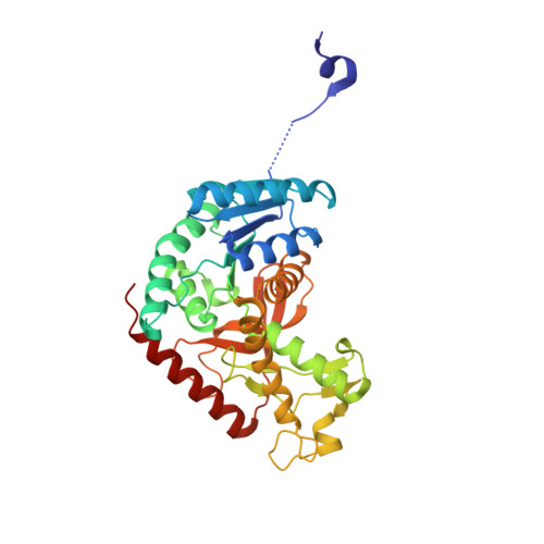
    Lactate dehydrogenase A (LDHA) is frequently overexpressed in tumors, thereby sustaining high glycolysis rates, tumor growth, and chemoresistance. High-throughput screening resulted in the identification of phthalimide and dibenzofuran derivatives as novel lactate dehydrogenase inhibitors, selectively inhibiting the activity of the LDHA isoenzyme. Cocrystallization experiments confirmed target engagement in addition to demonstrating binding to a novel allosteric binding site present in all four LDHA subunits of the LDH5 homotetramer.


  • Organizational Affiliation
    • Bayer AG, Pharmaceuticals, R&D, Müllerstrasse 178, 13342 Berlin, Germany.

Macromolecule Content 

  • Total Structure Weight: 298.14 kDa 
  • Atom Count: 20,754 
  • Modeled Residue Count: 2,624 
  • Deposited Residue Count: 2,656 
  • Unique protein chains: 1

Macromolecules

Find similar proteins by:|  3D Structure
Entity ID: 1
MoleculeChains  Sequence LengthOrganismDetailsImage
L-lactate dehydrogenase A chain
A, B, C, D, E
A, B, C, D, E, F, G, H
332Homo sapiensMutation(s): 0 
Gene Names: LDHAPIG19
EC: 1.1.1.27
UniProt & NIH Common Fund Data Resources
Find proteins for P00338 (Homo sapiens)
Explore P00338 
Go to UniProtKB:  P00338
PHAROS:  P00338
GTEx:  ENSG00000134333 
Entity Groups
Sequence Clusters30% Identity50% Identity70% Identity90% Identity95% Identity100% Identity
UniProt GroupP00338
Sequence Annotations
Expand
Reference Sequence

Small Molecules

Ligands 2 Unique
IDChains Name / Formula / InChI Key2D Diagram3D Interactions
NAI

Query on NAI


Download:Ideal Coordinates CCD File
J [auth A],
O [auth E]
1,4-DIHYDRONICOTINAMIDE ADENINE DINUCLEOTIDE
C21 H29 N7 O14 P2
BOPGDPNILDQYTO-NNYOXOHSSA-N
L5N
(Subject of Investigation/LOI)

Query on L5N


Download:Ideal Coordinates CCD File
I [auth A]
K [auth B]
L [auth C]
M [auth D]
N [auth E]
I [auth A],
K [auth B],
L [auth C],
M [auth D],
N [auth E],
P [auth F],
Q [auth G],
R [auth H]
4-[[4-[(5-chloranylthiophen-2-yl)carbonylamino]-1,3-bis(oxidanylidene)isoindol-2-yl]methyl]benzoic acid
C21 H13 Cl N2 O5 S
IOFCJSIYHLICRV-UHFFFAOYSA-N

Experimental Data & Validation

Experimental Data

  • Method: X-RAY DIFFRACTION
  • Resolution: 2.91 Å
  • R-Value Free:  0.244 (Depositor), 0.244 (DCC) 
  • R-Value Work:  0.207 (Depositor), 0.211 (DCC) 
  • R-Value Observed: 0.208 (Depositor) 
Space Group: P 21 21 21
Unit Cell:
Length ( Å )Angle ( ˚ )
a = 78.445α = 90
b = 143.853β = 90
c = 311.225γ = 90
Software Package:
Software NamePurpose
REFMACrefinement
XDSdata reduction
XSCALEdata scaling
PHASERphasing
Cootmodel building

Structure Validation

View Full Validation Report


Ligand Structure Quality Assessment 


Entry History 

Deposition Data

Revision History  (Full details and data files)

  • Version 1.0: 2020-06-10
    Type: Initial release
  • Version 1.1: 2020-07-01
    Changes: Database references
  • Version 1.2: 2024-01-24
    Changes: Data collection, Database references, Refinement description