6SHC | pdb_00006shc

Crystal structure of human IRE1 luminal domain Q105C


Experimental Data Snapshot

  • Method: X-RAY DIFFRACTION
  • Resolution: 3.55 Å
  • R-Value Free: 
    0.332 (Depositor), 0.329 (DCC) 
  • R-Value Work: 
    0.323 (Depositor), 0.332 (DCC) 
  • R-Value Observed: 
    0.324 (Depositor) 

Starting Model: experimental
View more details

wwPDB Validation 3D Report Full Report

Validation slider image for 6SHC

This is version 1.2 of the entry. See complete history

Literature

Unstructured regions in IRE1 alpha specify BiP-mediated destabilisation of the luminal domain dimer and repression of the UPR.

Amin-Wetzel, N.Neidhardt, L.Yan, Y.Mayer, M.P.Ron, D.

(2019) Elife 8

  • DOI: https://doi.org/10.7554/eLife.50793
  • Primary Citation Related Structures: 
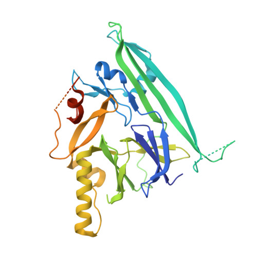
    6SHC

  • PubMed Abstract: 

    Coupling of endoplasmic reticulum (ER) stress to dimerisation-dependent activation of the UPR transducer IRE1 is incompletely understood. Whilst the luminal co-chaperone ERdj4 promotes a complex between the Hsp70 BiP and IRE1's stress-sensing luminal domain (IRE1 LD ) that favours the latter's monomeric inactive state and loss of ERdj4 de-represses IRE1, evidence linking these cellular and in vitro observations is presently lacking. We report that enforced loading of endogenous BiP onto endogenous IRE1α repressed UPR signalling in CHO cells and deletions in the IRE1α locus that de-repressed the UPR in cells, encode flexible regions of IRE1 LD that mediated BiP-induced monomerisation in vitro. Changes in the hydrogen exchange mass spectrometry profile of IRE1 LD induced by ERdj4 and BiP confirmed monomerisation and were consistent with active destabilisation of the IRE1 LD dimer. Together, these observations support a competition model whereby waning ER stress passively partitions ERdj4 and BiP to IRE1 LD to initiate active repression of UPR signalling.


  • Organizational Affiliation
    • Cambridge Institute for Medical Research (CIMR), University of Cambridge, Cambridge, United Kingdom.

Macromolecule Content 

  • Total Structure Weight: 41.01 kDa 
  • Atom Count: 1,784 
  • Modeled Residue Count: 273 
  • Deposited Residue Count: 367 
  • Unique protein chains: 1

Macromolecules

Find similar proteins by:|  3D Structure
Entity ID: 1
MoleculeChains  Sequence LengthOrganismDetailsImage
Serine/threonine-protein kinase/endoribonuclease IRE1367Homo sapiensMutation(s): 4 
Gene Names: ERN1IRE1
EC: 2.7.11.1 (PDB Primary Data), 3.1.26 (PDB Primary Data)
UniProt & NIH Common Fund Data Resources
Find proteins for O75460 (Homo sapiens)
Explore O75460 
Go to UniProtKB:  O75460
PHAROS:  O75460
GTEx:  ENSG00000178607 
Entity Groups
Sequence Clusters30% Identity50% Identity70% Identity90% Identity95% Identity100% Identity
UniProt GroupO75460
Sequence Annotations
Expand
Reference Sequence

Experimental Data & Validation

Experimental Data

  • Method: X-RAY DIFFRACTION
  • Resolution: 3.55 Å
  • R-Value Free:  0.332 (Depositor), 0.329 (DCC) 
  • R-Value Work:  0.323 (Depositor), 0.332 (DCC) 
  • R-Value Observed: 0.324 (Depositor) 
Space Group: P 65 2 2
Unit Cell:
Length ( Å )Angle ( ˚ )
a = 182.772α = 90
b = 182.772β = 90
c = 68.449γ = 120
Software Package:
Software NamePurpose
PHENIXrefinement
REFMACrefinement
DIALSdata reduction
Aimlessdata scaling
PHASERphasing

Structure Validation

View Full Validation Report


Entry History 

& Funding Information

Deposition Data


Funding OrganizationLocationGrant Number
Wellcome TrustUnited KingdomWellcome 200848/Z/16/Z
Wellcome TrustUnited KingdomWellcome 100140

Revision History  (Full details and data files)

  • Version 1.0: 2020-01-08
    Type: Initial release
  • Version 1.1: 2024-01-24
    Changes: Data collection, Database references, Refinement description
  • Version 1.2: 2024-10-23
    Changes: Structure summary