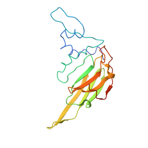
6T1R

Pseudo-atomic model of a 16-mer assembly of reduced recombinant human alphaA-crystallin (non domain swapped configuration)


Experimental Data Snapshot

  • Method: ELECTRON MICROSCOPY
  • Resolution: 9.80 Å
  • Aggregation State: PARTICLE 
  • Reconstruction Method: SINGLE PARTICLE 

Starting Model: experimental
View more details

wwPDB Validation   3D Report Full Report


This is version 1.2 of the entry. See complete history


Literature

The structure and oxidation of the eye lens chaperone alpha A-crystallin.

Kaiser, C.J.O.Peters, C.Schmid, P.W.N.Stavropoulou, M.Zou, J.Dahiya, V.Mymrikov, E.V.Rockel, B.Asami, S.Haslbeck, M.Rappsilber, J.Reif, B.Zacharias, M.Buchner, J.Weinkauf, S.

(2019) Nat Struct Mol Biol 26: 1141-1150

  • DOI: https://doi.org/10.1038/s41594-019-0332-9
  • Primary Citation of Related Structures:  
    6T1R

  • PubMed Abstract: 

    The small heat shock protein αA-crystallin is a molecular chaperone important for the optical properties of the vertebrate eye lens. It forms heterogeneous oligomeric ensembles. We determined the structures of human αA-crystallin oligomers by combining cryo-electron microscopy, cross-linking/mass spectrometry, NMR spectroscopy and molecular modeling. The different oligomers can be interconverted by the addition or subtraction of tetramers, leading to mainly 12-, 16- and 20-meric assemblies in which interactions between N-terminal regions are important. Cross-dimer domain-swapping of the C-terminal region is a determinant of αA-crystallin heterogeneity. Human αA-crystallin contains two cysteines, which can form an intramolecular disulfide in vivo. Oxidation in vitro requires conformational changes and oligomer dissociation. The oxidized oligomers, which are larger than reduced αA-crystallin and destabilized against unfolding, are active chaperones and can transfer the disulfide to destabilized substrate proteins. The insight into the structure and function of αA-crystallin provides a basis for understanding its role in the eye lens.


  • Organizational Affiliation

    Center for Integrated Protein Science Munich at the Department Chemie, Technische Universität München, Garching, Germany.


Macromolecules
Find similar proteins by:  (by identity cutoff)  |  3D Structure
Entity ID: 1
MoleculeChains Sequence LengthOrganismDetailsImage
Alpha-crystallin A chain
A, B, C, D, E
A, B, C, D, E, F, G, H, I, J, K, L, M, N, O, P
173Homo sapiensMutation(s): 0 
Gene Names: CRYAACRYA1HSPB4
UniProt & NIH Common Fund Data Resources
Find proteins for P02489 (Homo sapiens)
Explore P02489 
Go to UniProtKB:  P02489
PHAROS:  P02489
GTEx:  ENSG00000160202 
Entity Groups  
Sequence Clusters30% Identity50% Identity70% Identity90% Identity95% Identity100% Identity
UniProt GroupP02489
Sequence Annotations
Expand
  • Reference Sequence
Experimental Data & Validation

Experimental Data

  • Method: ELECTRON MICROSCOPY
  • Resolution: 9.80 Å
  • Aggregation State: PARTICLE 
  • Reconstruction Method: SINGLE PARTICLE 
EM Software:
TaskSoftware PackageVersion
RECONSTRUCTIONIMAGIC5
MODEL REFINEMENTAmber16

Structure Validation

View Full Validation Report


Entry History & Funding Information

Deposition Data


Funding OrganizationLocationGrant Number
German Research FoundationGermanyCRC 1035
German Research FoundationGermanyEXC 114

Revision History  (Full details and data files)

  • Version 1.0: 2019-12-11
    Type: Initial release
  • Version 1.1: 2019-12-18
    Changes: Database references
  • Version 1.2: 2024-05-15
    Changes: Data collection, Database references, Refinement description