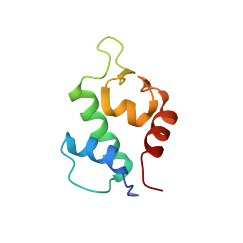
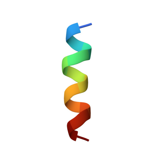
6T2D

Multicomponent Peptide Stapling as a Diversity-Driven Tool for the Development of Inhibitors of Protein-Protein Interactions


Experimental Data Snapshot

  • Method: X-RAY DIFFRACTION
  • Resolution: 1.80 Å
  • R-Value Free: 0.226 
  • R-Value Work: 0.132 
  • R-Value Observed: 0.136 

Starting Model: experimental
View more details

wwPDB Validation   3D Report Full Report


Ligand Structure Quality Assessment 


This is version 1.3 of the entry. See complete history


Literature

Multicomponent Peptide Stapling as a Diversity-Driven Tool for the Development of Inhibitors of Protein-Protein Interactions.

Ricardo, M.G.Ali, A.M.Plewka, J.Surmiak, E.Labuzek, B.Neochoritis, C.G.Atmaj, J.Skalniak, L.Zhang, R.Holak, T.A.Groves, M.Rivera, D.G.Domling, A.

(2020) Angew Chem Int Ed Engl 59: 5235-5241

  • DOI: https://doi.org/10.1002/anie.201916257
  • Primary Citation of Related Structures:  
    6T2D, 6T2E, 6T2F

  • PubMed Abstract: 

    Stapled peptides are chemical entities in-between biologics and small molecules, which have proven to be the solution to high affinity protein-protein interaction antagonism, while keeping control over pharmacological performance such as stability and membrane penetration. We demonstrate that the multicomponent reaction-based stapling is an effective strategy for the development of α-helical peptides with highly potent dual antagonistic action of MDM2 and MDMX binding p53. Such a potent inhibitory activity of p53-MDM2/X interactions was assessed by fluorescence polarization, microscale thermophoresis, and 2D NMR, while several cocrystal structures with MDM2 were obtained. This MCR stapling protocol proved efficient and versatile in terms of diversity generation at the staple, as evidenced by the incorporation of both exo- and endo-cyclic hydrophobic moieties at the side chain cross-linkers. The interaction of the Ugi-staple fragments with the target protein was demonstrated by crystallography.


  • Organizational Affiliation

    Faculty of Chemistry, Center for Natural Product Research, University of Havana, Cuba.


Macromolecules
Find similar proteins by:  (by identity cutoff)  |  3D Structure
Entity ID: 1
MoleculeChains Sequence LengthOrganismDetailsImage
E3 ubiquitin-protein ligase Mdm286Homo sapiensMutation(s): 1 
Gene Names: MDM2
EC: 2.3.2.27
UniProt & NIH Common Fund Data Resources
Find proteins for Q00987 (Homo sapiens)
Explore Q00987 
Go to UniProtKB:  Q00987
PHAROS:  Q00987
GTEx:  ENSG00000135679 
Entity Groups  
Sequence Clusters30% Identity50% Identity70% Identity90% Identity95% Identity100% Identity
UniProt GroupQ00987
Sequence Annotations
Expand
  • Reference Sequence

Find similar proteins by:  Sequence   |   3D Structure  

Entity ID: 2
MoleculeChains Sequence LengthOrganismDetailsImage
Stapled peptide GAR300-Gp14synthetic constructMutation(s): 0 
Entity Groups  
Sequence Clusters30% Identity50% Identity70% Identity90% Identity95% Identity100% Identity
Sequence Annotations
Expand
  • Reference Sequence
Small Molecules
Ligands 3 Unique
IDChains Name / Formula / InChI Key2D Diagram3D Interactions
M9E (Subject of Investigation/LOI)
Query on M9E

Download Ideal Coordinates CCD File 
E [auth B]2-(methylamino)-~{N}-[[4-[[2-(methylamino)ethanoylamino]methyl]phenyl]methyl]ethanamide
C14 H22 N4 O2
RZNHDMVQBYDUNO-UHFFFAOYSA-N
EPE
Query on EPE

Download Ideal Coordinates CCD File 
C [auth A]4-(2-HYDROXYETHYL)-1-PIPERAZINE ETHANESULFONIC ACID
C8 H18 N2 O4 S
JKMHFZQWWAIEOD-UHFFFAOYSA-N
DMS
Query on DMS

Download Ideal Coordinates CCD File 
D [auth A]DIMETHYL SULFOXIDE
C2 H6 O S
IAZDPXIOMUYVGZ-UHFFFAOYSA-N
Experimental Data & Validation

Experimental Data

  • Method: X-RAY DIFFRACTION
  • Resolution: 1.80 Å
  • R-Value Free: 0.226 
  • R-Value Work: 0.132 
  • R-Value Observed: 0.136 
  • Space Group: P 31 2 1
Unit Cell:
Length ( Å )Angle ( ˚ )
a = 41.017α = 90
b = 41.017β = 90
c = 104.995γ = 120
Software Package:
Software NamePurpose
REFMACrefinement
XDSdata reduction
Aimlessdata scaling
PDB_EXTRACTdata extraction
PHASERphasing

Structure Validation

View Full Validation Report


Ligand Structure Quality Assessment 


Entry History 

Deposition Data

Revision History  (Full details and data files)

  • Version 1.0: 2020-01-29
    Type: Initial release
  • Version 1.1: 2020-03-25
    Changes: Database references
  • Version 1.2: 2024-01-24
    Changes: Data collection, Database references, Refinement description
  • Version 1.3: 2024-10-23
    Changes: Structure summary