6T41

CDK8/Cyclin C in complex with N-(4-chlorobenzyl)isoquinolin-4-amine


Experimental Data Snapshot

  • Method: X-RAY DIFFRACTION
  • Resolution: 2.45 Å
  • R-Value Free: 0.256 
  • R-Value Work: 0.183 
  • R-Value Observed: 0.185 

wwPDB Validation   3D Report Full Report


Ligand Structure Quality Assessment 


This is version 1.3 of the entry. See complete history


Literature

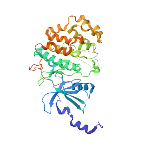
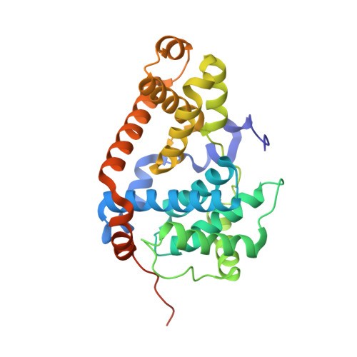
A precisely positioned MED12 activation helix stimulates CDK8 kinase activity.

Klatt, F.Leitner, A.Kim, I.V.Ho-Xuan, H.Schneider, E.V.Langhammer, F.Weinmann, R.Muller, M.R.Huber, R.Meister, G.Kuhn, C.D.

(2020) Proc Natl Acad Sci U S A 117: 2894-2905

  • DOI: https://doi.org/10.1073/pnas.1917635117
  • Primary Citation of Related Structures:  
    6T41

  • PubMed Abstract: 

    The Mediator kinase module regulates eukaryotic transcription by phosphorylating transcription-related targets and by modulating the association of Mediator and RNA polymerase II. The activity of its catalytic core, cyclin-dependent kinase 8 (CDK8), is controlled by Cyclin C and regulatory subunit MED12, with its deregulation contributing to numerous malignancies. Here, we combine in vitro biochemistry, cross-linking coupled to mass spectrometry, and in vivo studies to describe the binding location of the N-terminal segment of MED12 on the CDK8/Cyclin C complex and to gain mechanistic insights into the activation of CDK8 by MED12. Our data demonstrate that the N-terminal portion of MED12 wraps around CDK8, whereby it positions an "activation helix" close to the T-loop of CDK8 for its activation. Intriguingly, mutations in the activation helix that are frequently found in cancers do not diminish the affinity of MED12 for CDK8, yet likely alter the exact positioning of the activation helix. Furthermore, we find the transcriptome-wide gene-expression changes in human cells that result from a mutation in the MED12 activation helix to correlate with deregulated genes in breast and colon cancer. Finally, functional assays in the presence of kinase inhibitors reveal that binding of MED12 remodels the active site of CDK8 and thereby precludes the inhibition of ternary CDK8 complexes by type II kinase inhibitors. Taken together, our results not only allow us to propose a revised model of how CDK8 activity is regulated by MED12, but also offer a path forward in developing small molecules that target CDK8 in its MED12-bound form.


  • Organizational Affiliation

    Gene Regulation by Non-Coding RNA, Elite Network of Bavaria and University of Bayreuth, 95447 Bayreuth, Germany.


Macromolecules
Find similar proteins by:  (by identity cutoff)  |  3D Structure
Entity ID: 1
MoleculeChains Sequence LengthOrganismDetailsImage
Cyclin-dependent kinase 8407Homo sapiensMutation(s): 0 
Gene Names: CDK8
EC: 2.7.11.22 (PDB Primary Data), 2.7.11.23 (PDB Primary Data)
UniProt & NIH Common Fund Data Resources
Find proteins for P49336 (Homo sapiens)
Explore P49336 
Go to UniProtKB:  P49336
PHAROS:  P49336
GTEx:  ENSG00000132964 
Entity Groups  
Sequence Clusters30% Identity50% Identity70% Identity90% Identity95% Identity100% Identity
UniProt GroupP49336
Sequence Annotations
Expand
  • Reference Sequence
Find similar proteins by:  (by identity cutoff)  |  3D Structure
Entity ID: 2
MoleculeChains Sequence LengthOrganismDetailsImage
Cyclin-C286Homo sapiensMutation(s): 0 
Gene Names: CCNC
UniProt & NIH Common Fund Data Resources
Find proteins for P24863 (Homo sapiens)
Explore P24863 
Go to UniProtKB:  P24863
PHAROS:  P24863
GTEx:  ENSG00000112237 
Entity Groups  
Sequence Clusters30% Identity50% Identity70% Identity90% Identity95% Identity100% Identity
UniProt GroupP24863
Sequence Annotations
Expand
  • Reference Sequence
Experimental Data & Validation

Experimental Data

  • Method: X-RAY DIFFRACTION
  • Resolution: 2.45 Å
  • R-Value Free: 0.256 
  • R-Value Work: 0.183 
  • R-Value Observed: 0.185 
  • Space Group: P 21 21 21
Unit Cell:
Length ( Å )Angle ( ˚ )
a = 70.969α = 90
b = 70.687β = 90
c = 170.253γ = 90
Software Package:
Software NamePurpose
PHENIXrefinement
XSCALEdata scaling
PDB_EXTRACTdata extraction
XDSdata reduction
MOLREPphasing

Structure Validation

View Full Validation Report


Ligand Structure Quality Assessment 


Entry History 

Deposition Data

Revision History  (Full details and data files)

  • Version 1.0: 2020-01-01
    Type: Initial release
  • Version 1.1: 2020-01-22
    Changes: Database references
  • Version 1.2: 2020-02-19
    Changes: Database references
  • Version 1.3: 2024-01-24
    Changes: Data collection, Database references, Refinement description