6TDQ | pdb_00006tdq

Crystal structure of the disulfide engineered HLA-A0201 molecule in complex with one GM dipeptide in the A pocket and one GM dipeptide in the F pocket.


Experimental Data Snapshot

  • Method: X-RAY DIFFRACTION
  • Resolution: 1.60 Å
  • R-Value Free: 
    0.214 (Depositor), 0.221 (DCC) 
  • R-Value Work: 
    0.169 (Depositor), 0.183 (DCC) 
  • R-Value Observed: 
    0.171 (Depositor) 

Starting Model: experimental
View more details

wwPDB Validation 3D Report Full Report

Validation slider image for 6TDQ

Ligand Structure Quality Assessment 


This is version 1.2 of the entry. See complete history

Literature

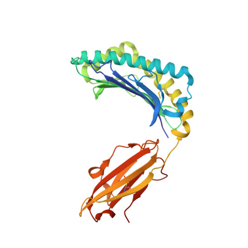
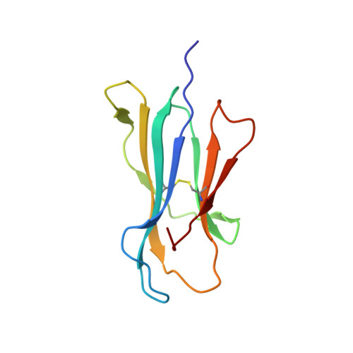
Structures of peptide-free and partially loaded MHC class I molecules reveal mechanisms of peptide selection.

Anjanappa, R.Garcia-Alai, M.Kopicki, J.D.Lockhauserbaumer, J.Aboelmagd, M.Hinrichs, J.Nemtanu, I.M.Uetrecht, C.Zacharias, M.Springer, S.Meijers, R.

(2020) Nat Commun 11: 1314-1314

  • DOI: https://doi.org/10.1038/s41467-020-14862-4
  • Primary Citation Related Structures: 
    6TDO, 6TDP, 6TDQ, 6TDR, 6TDS

  • PubMed Abstract: 

    Major Histocompatibility Complex (MHC) class I molecules selectively bind peptides for presentation to cytotoxic T cells. The peptide-free state of these molecules is not well understood. Here, we characterize a disulfide-stabilized version of the human class I molecule HLA-A*02:01 that is stable in the absence of peptide and can readily exchange cognate peptides. We present X-ray crystal structures of the peptide-free state of HLA-A*02:01, together with structures that have dipeptides bound in the A and F pockets. These structural snapshots reveal that the amino acid side chains lining the binding pockets switch in a coordinated fashion between a peptide-free unlocked state and a peptide-bound locked state. Molecular dynamics simulations suggest that the opening and closing of the F pocket affects peptide ligand conformations in adjacent binding pockets. We propose that peptide binding is co-determined by synergy between the binding pockets of the MHC molecule.


  • Organizational Affiliation
    • Department of Life Sciences and Chemistry, Jacobs University Bremen, Bremen, Germany.

Macromolecule Content 

  • Total Structure Weight: 89.1 kDa 
  • Atom Count: 7,718 
  • Modeled Residue Count: 750 
  • Deposited Residue Count: 752 
  • Unique protein chains: 2

Macromolecules

Find similar proteins by:|  3D Structure
Entity ID: 1
MoleculeChains  Sequence LengthOrganismDetailsImage
MHC class I antigen
A, C
276Homo sapiensMutation(s): 0 
Gene Names: HLA-A
UniProt
Find proteins for F6IQS1 (Homo sapiens)
Explore F6IQS1 
Go to UniProtKB:  F6IQS1
Entity Groups
Sequence Clusters30% Identity50% Identity70% Identity90% Identity95% Identity100% Identity
UniProt GroupF6IQS1
Sequence Annotations
Expand
Reference Sequence
Find similar proteins by:|  3D Structure
Entity ID: 2
MoleculeChains  Sequence LengthOrganismDetailsImage
Beta-2-microglobulin
B, D
100Homo sapiensMutation(s): 0 
Gene Names: B2MCDABP0092HDCMA22P
UniProt & NIH Common Fund Data Resources
Find proteins for P61769 (Homo sapiens)
Explore P61769 
Go to UniProtKB:  P61769
PHAROS:  P61769
GTEx:  ENSG00000166710 
Entity Groups
Sequence Clusters30% Identity50% Identity70% Identity90% Identity95% Identity100% Identity
UniProt GroupP61769
Sequence Annotations
Expand
Reference Sequence

Small Molecules

Ligands 4 Unique
IDChains Name / Formula / InChI Key2D Diagram3D Interactions
MET

Query on MET


Download:Ideal Coordinates CCD File
H [auth A],
J [auth A],
S [auth C],
U [auth C]
METHIONINE
C5 H11 N O2 S
FFEARJCKVFRZRR-BYPYZUCNSA-N
GLY

Query on GLY


Download:Ideal Coordinates CCD File
G [auth A],
I [auth A],
R [auth C],
T [auth C]
GLYCINE
C2 H5 N O2
DHMQDGOQFOQNFH-UHFFFAOYSA-N
EDO
(Subject of Investigation/LOI)

Query on EDO


Download:Ideal Coordinates CCD File
E [auth A]
F [auth A]
L [auth B]
M [auth B]
N [auth B]
E [auth A],
F [auth A],
L [auth B],
M [auth B],
N [auth B],
O [auth C],
P [auth C],
Q [auth C],
V [auth D]
1,2-ETHANEDIOL
C2 H6 O2
LYCAIKOWRPUZTN-UHFFFAOYSA-N
CL
(Subject of Investigation/LOI)

Query on CL


Download:Ideal Coordinates CCD File
K [auth A]CHLORIDE ION
Cl
VEXZGXHMUGYJMC-UHFFFAOYSA-M

Experimental Data & Validation

Experimental Data

  • Method: X-RAY DIFFRACTION
  • Resolution: 1.60 Å
  • R-Value Free:  0.214 (Depositor), 0.221 (DCC) 
  • R-Value Work:  0.169 (Depositor), 0.183 (DCC) 
  • R-Value Observed: 0.171 (Depositor) 
Space Group: P 1 21 1
Unit Cell:
Length ( Å )Angle ( ˚ )
a = 58.643α = 90
b = 84.124β = 90
c = 83.716γ = 90
Software Package:
Software NamePurpose
REFMACrefinement
PDB_EXTRACTdata extraction
XDSdata reduction
Aimlessdata scaling
MOLREPphasing

Structure Validation

View Full Validation Report


Ligand Structure Quality Assessment 


Entry History 

& Funding Information

Deposition Data


Funding OrganizationLocationGrant Number
European CommissionGermany653706

Revision History  (Full details and data files)

  • Version 1.0: 2020-03-25
    Type: Initial release
  • Version 1.1: 2024-01-24
    Changes: Data collection, Database references, Refinement description
  • Version 1.2: 2024-10-16
    Changes: Structure summary