6TF7 | pdb_00006tf7

Human galectin-3c in complex with a galactose derivative


Experimental Data Snapshot

  • Method: X-RAY DIFFRACTION
  • Resolution: 1.40 Å
  • R-Value Free: 
    0.187 (Depositor), 0.188 (DCC) 
  • R-Value Work: 
    0.135 (Depositor) 
  • R-Value Observed: 
    0.137 (Depositor) 

Starting Model: experimental
View more details

wwPDB Validation 3D Report Full Report

Validation slider image for 6TF7

Ligand Structure Quality Assessment 


This is version 1.2 of the entry. See complete history

Literature

3-Substituted 1-Naphthamidomethyl-C-galactosyls Interact with Two Unique Sub-sites for High-Affinity and High-Selectivity Inhibition of Galectin-3.

Dahlqvist, A.Mandal, S.Peterson, K.Hakansson, M.Logan, D.T.Zetterberg, F.R.Leffler, H.Nilsson, U.J.

(2019) Molecules 24

  • DOI: https://doi.org/10.3390/molecules24244554
  • Primary Citation Related Structures: 
    6TF6, 6TF7

  • PubMed Abstract: 

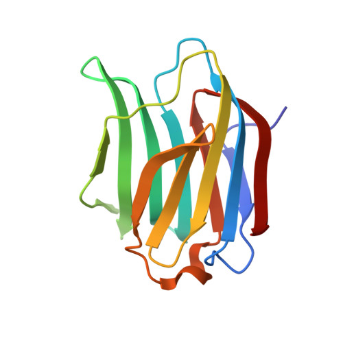
    The galectins are a family of galactose-binding proteins playing key roles in inflammatory processes and cancer. However, they are structurally very closely related, and discovery of highly selective inhibitors is challenging. In this work, we report the design of novel inhibitors binding to a subsite unique to galectin-3, which confers both high selectivity and affinity towards galectin-3. Olefin cross metathesis between allyl β-C-galactopyranosyl and 1-vinylnaphthalenes or acylation of aminomethyl β-C-galactopyranosyl with 1-naphthoic acid derivatives gave C-galactopyranosyls carrying 1-naphthamide structural elements that interacted favorably with a galectin-3 unique subsite according to molecular modeling and X-ray structural analysis of two inhibitor-galectin-3 complexes. Affinities were down to sub-µM and selectivities over galectin-1, 2, 4 N -terminal domain, 4 C-terminal domain, 7, 8 N -terminal domain, 9 N -terminal domain, and 9 C-terminal domain were high. These results show that high affinity and selectivity for a single galectin can be achieved by targeting unique subsites, which holds promise for further development of small and selective galectin inhibitors.


  • Organizational Affiliation
    • Centre for Analysis and Synthesis, Department of Chemistry, Lund University, POB124, SE-22100 Lund, Sweden.

Macromolecule Content 

  • Total Structure Weight: 16.45 kDa 
  • Atom Count: 1,468 
  • Modeled Residue Count: 138 
  • Deposited Residue Count: 138 
  • Unique protein chains: 1

Macromolecules

Find similar proteins by:|  3D Structure
Entity ID: 1
MoleculeChains  Sequence LengthOrganismDetailsImage
Galectin-3138Homo sapiensMutation(s): 0 
Gene Names: LGALS3MAC2
UniProt & NIH Common Fund Data Resources
Find proteins for P17931 (Homo sapiens)
Explore P17931 
Go to UniProtKB:  P17931
PHAROS:  P17931
GTEx:  ENSG00000131981 
Entity Groups
Sequence Clusters30% Identity50% Identity70% Identity90% Identity95% Identity100% Identity
UniProt GroupP17931
Sequence Annotations
Expand
Reference Sequence

Experimental Data & Validation

Experimental Data

  • Method: X-RAY DIFFRACTION
  • Resolution: 1.40 Å
  • R-Value Free:  0.187 (Depositor), 0.188 (DCC) 
  • R-Value Work:  0.135 (Depositor) 
  • R-Value Observed: 0.137 (Depositor) 
Space Group: P 21 21 21
Unit Cell:
Length ( Å )Angle ( ˚ )
a = 36.14α = 90
b = 58.16β = 90
c = 62.84γ = 90
Software Package:
Software NamePurpose
XDSdata reduction
XSCALEdata scaling
REFMACrefinement
PDB_EXTRACTdata extraction
REFMACphasing

Structure Validation

View Full Validation Report


Ligand Structure Quality Assessment 


Entry History 

& Funding Information

Deposition Data


Funding OrganizationLocationGrant Number
Swedish Research CouncilSweden621-2012-2978

Revision History  (Full details and data files)

  • Version 1.0: 2020-11-18
    Type: Initial release
  • Version 1.1: 2021-12-01
    Changes: Advisory, Database references
  • Version 1.2: 2024-01-24
    Changes: Data collection, Refinement description