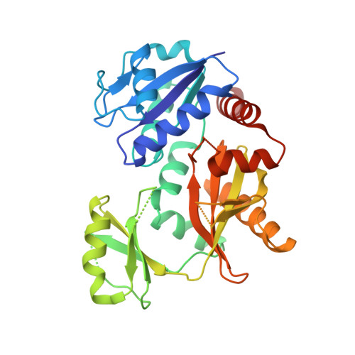
6U1I

Thermus thermophilus D-alanine-D-alanine ligase in complex with ADP, phosphorylated D-cycloserine, Mg2+ and K+


Experimental Data Snapshot

  • Method: X-RAY DIFFRACTION
  • Resolution: 2.30 Å
  • R-Value Free: 0.280 
  • R-Value Work: 0.238 
  • R-Value Observed: 0.240 

Starting Model: experimental
View more details

wwPDB Validation   3D Report Full Report


Ligand Structure Quality Assessment 


This is version 1.2 of the entry. See complete history


Literature

d-Alanine-d-alanine ligase as a model for the activation of ATP-grasp enzymes by monovalent cations.

Pederick, J.L.Thompson, A.P.Bell, S.G.Bruning, J.B.

(2020) J Biol Chem 295: 7894-7904

  • DOI: https://doi.org/10.1074/jbc.RA120.012936
  • Primary Citation of Related Structures:  
    6U1C, 6U1D, 6U1E, 6U1F, 6U1G, 6U1H, 6U1I, 6U1J, 6U1K

  • PubMed Abstract: 

    The ATP-grasp superfamily of enzymes shares an atypical nucleotide-binding site known as the ATP-grasp fold. These enzymes are involved in many biological pathways in all domains of life. One ATP-grasp enzyme, d-alanine-d-alanine ligase (Ddl), catalyzes ATP-dependent formation of the d-alanyl-d-alanine dipeptide essential for bacterial cell wall biosynthesis and is therefore an important antibiotic drug target. Ddl is activated by the monovalent cation (MVC) K + , but despite its clinical relevance and decades of research, how this activation occurs has not been elucidated. We demonstrate here that activating MVCs bind adjacent to the active site of Ddl from Thermus thermophilus and used a combined biochemical and structural approach to characterize MVC activation. We found that Tt Ddl is a type II MVC-activated enzyme, retaining activity in the absence of MVCs. However, the efficiency of Tt Ddl increased ∼20-fold in the presence of activating MVCs, and it was maximally activated by K + and Rb + ions. A strict dependence on ionic radius of the MVC was observed, with Li + and Na + providing little to no Tt Ddl activation. To understand the mechanism of MVC activation, we solved crystal structures of Tt Ddl representing distinct catalytic stages in complex with K + , Rb + , or Cs + Comparison of these structures with apo Tt Ddl revealed no evident conformational change on MVC binding. Of note, the identified MVC binding site is structurally conserved within the ATP-grasp superfamily. We propose that MVCs activate Ddl by altering the charge distribution of its active site. These findings provide insight into the catalytic mechanism of ATP-grasp enzymes.


  • Organizational Affiliation

    Institute for Photonics and Advanced Sensing, School of Biological Sciences, The University of Adelaide, Adelaide, South Australia, Australia.


Macromolecules
Find similar proteins by:  (by identity cutoff)  |  3D Structure
Entity ID: 1
MoleculeChains Sequence LengthOrganismDetailsImage
D-alanine--D-alanine ligase
A, B, C, D
327Thermus thermophilus HB8Mutation(s): 0 
Gene Names: ddlTTHA1587
EC: 6.3.2.4
UniProt
Find proteins for Q5SHZ3 (Thermus thermophilus (strain ATCC 27634 / DSM 579 / HB8))
Explore Q5SHZ3 
Go to UniProtKB:  Q5SHZ3
Entity Groups  
Sequence Clusters30% Identity50% Identity70% Identity90% Identity95% Identity100% Identity
UniProt GroupQ5SHZ3
Sequence Annotations
Expand
  • Reference Sequence
Small Molecules
Ligands 4 Unique
IDChains Name / Formula / InChI Key2D Diagram3D Interactions
ADP
Query on ADP

Download Ideal Coordinates CCD File 
E [auth A],
I [auth C]
ADENOSINE-5'-DIPHOSPHATE
C10 H15 N5 O10 P2
XTWYTFMLZFPYCI-KQYNXXCUSA-N
DS0
Query on DS0

Download Ideal Coordinates CCD File 
J [auth C][(4R)-4-azanyl-4,5-dihydro-1,2-oxazol-3-yl] dihydrogen phosphate
C3 H7 N2 O5 P
SFMNESRSKJTEMJ-UWTATZPHSA-N
K
Query on K

Download Ideal Coordinates CCD File 
H [auth C]POTASSIUM ION
K
NPYPAHLBTDXSSS-UHFFFAOYSA-N
MG
Query on MG

Download Ideal Coordinates CCD File 
F [auth C],
G [auth C]
MAGNESIUM ION
Mg
JLVVSXFLKOJNIY-UHFFFAOYSA-N
Experimental Data & Validation

Experimental Data

  • Method: X-RAY DIFFRACTION
  • Resolution: 2.30 Å
  • R-Value Free: 0.280 
  • R-Value Work: 0.238 
  • R-Value Observed: 0.240 
  • Space Group: P 1
Unit Cell:
Length ( Å )Angle ( ˚ )
a = 55.369α = 69.5
b = 68.453β = 78.86
c = 91.747γ = 66.05
Software Package:
Software NamePurpose
PHENIXrefinement
XDSdata reduction
Aimlessdata scaling
PHASERphasing
PDB_EXTRACTdata extraction

Structure Validation

View Full Validation Report


Ligand Structure Quality Assessment 


Entry History 

Deposition Data

Revision History  (Full details and data files)

  • Version 1.0: 2020-05-06
    Type: Initial release
  • Version 1.1: 2020-06-17
    Changes: Database references
  • Version 1.2: 2023-10-11
    Changes: Data collection, Database references, Derived calculations, Refinement description