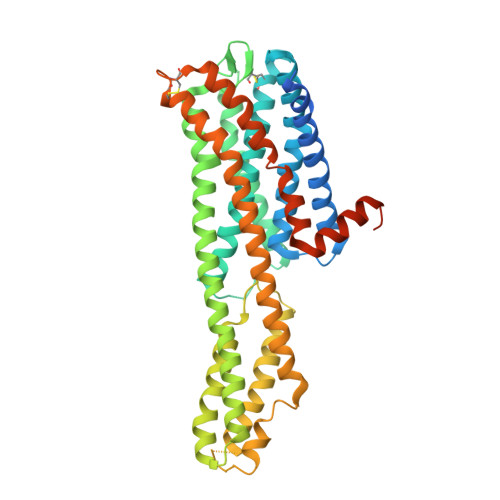
6WGT

Crystal structure of HTR2A with hallucinogenic agonist


Experimental Data Snapshot

  • Method: X-RAY DIFFRACTION
  • Resolution: 3.40 Å
  • R-Value Free: 0.305 
  • R-Value Work: 0.265 
  • R-Value Observed: 0.267 

Starting Model: experimental
View more details

wwPDB Validation   3D Report Full Report


Ligand Structure Quality Assessment 


This is version 1.3 of the entry. See complete history


Literature

Structure of a Hallucinogen-Activated Gq-Coupled 5-HT 2A Serotonin Receptor

Kim, K.L.Che, T.Panova, O.DiBerto, J.F.Lyu, J.Krumm, B.E.Wacker, D.Robertson, M.J.Seven, A.B.Nichols, D.E.Shoichet, B.K.Skiniotis, G.Roth, B.L.

(2020) Cell 182: 1574-1588


Macromolecules
Find similar proteins by:  (by identity cutoff)  |  3D Structure
Entity ID: 1
MoleculeChains Sequence LengthOrganismDetailsImage
5-hydroxytryptamine receptor 2A,Soluble cytochrome b562 fusion
A, B, C
448Homo sapiensEscherichia coliMutation(s): 0 
Gene Names: HTR2AHTR2cybC
Membrane Entity: Yes 
UniProt & NIH Common Fund Data Resources
Find proteins for P28223 (Homo sapiens)
Explore P28223 
Go to UniProtKB:  P28223
PHAROS:  P28223
GTEx:  ENSG00000102468 
Find proteins for P0ABE7 (Escherichia coli)
Explore P0ABE7 
Go to UniProtKB:  P0ABE7
Entity Groups  
Sequence Clusters30% Identity50% Identity70% Identity90% Identity95% Identity100% Identity
UniProt GroupsP28223P0ABE7
Sequence Annotations
Expand
  • Reference Sequence
Small Molecules
Ligands 6 Unique
IDChains Name / Formula / InChI Key2D Diagram3D Interactions
CLR
Query on CLR

Download Ideal Coordinates CCD File 
E [auth A],
I [auth A]
CHOLESTEROL
C27 H46 O
HVYWMOMLDIMFJA-DPAQBDIFSA-N
7LD (Subject of Investigation/LOI)
Query on 7LD

Download Ideal Coordinates CCD File 
D [auth A],
J [auth B],
N [auth C]
(8alpha)-N,N-diethyl-6-methyl-9,10-didehydroergoline-8-carboxamide
C20 H25 N3 O
VAYOSLLFUXYJDT-RDTXWAMCSA-N
OLA
Query on OLA

Download Ideal Coordinates CCD File 
F [auth A],
G [auth A],
K [auth B]
OLEIC ACID
C18 H34 O2
ZQPPMHVWECSIRJ-KTKRTIGZSA-N
1PE
Query on 1PE

Download Ideal Coordinates CCD File 
Q [auth C]PENTAETHYLENE GLYCOL
C10 H22 O6
JLFNLZLINWHATN-UHFFFAOYSA-N
PEG
Query on PEG

Download Ideal Coordinates CCD File 
H [auth A],
O [auth C]
DI(HYDROXYETHYL)ETHER
C4 H10 O3
MTHSVFCYNBDYFN-UHFFFAOYSA-N
PO4
Query on PO4

Download Ideal Coordinates CCD File 
L [auth B],
M [auth B],
P [auth C]
PHOSPHATE ION
O4 P
NBIIXXVUZAFLBC-UHFFFAOYSA-K
Binding Affinity Annotations 
IDSourceBinding Affinity
7LD BindingDB:  6WGT Ki: min: 0.76, max: 21.38 (nM) from 19 assay(s)
Kd: 21.17 (nM) from 1 assay(s)
EC50: min: 5.2, max: 15 (nM) from 5 assay(s)
Experimental Data & Validation

Experimental Data

  • Method: X-RAY DIFFRACTION
  • Resolution: 3.40 Å
  • R-Value Free: 0.305 
  • R-Value Work: 0.265 
  • R-Value Observed: 0.267 
  • Space Group: C 2 2 21
Unit Cell:
Length ( Å )Angle ( ˚ )
a = 81.64α = 90
b = 175.26β = 90
c = 280.67γ = 90
Software Package:
Software NamePurpose
PHENIXrefinement
Cootmodel building
XDSdata reduction
XDSdata scaling
PHENIXphasing

Structure Validation

View Full Validation Report


Ligand Structure Quality Assessment 


Entry History & Funding Information

Deposition Data


Funding OrganizationLocationGrant Number
National Institutes of Health/National Institute on Drug Abuse (NIH/NIDA)United StatesR37 DA045657
National Institutes of Health/National Institute of Mental Health (NIH/NIMH)United StatesRO1 MH112205

Revision History  (Full details and data files)

  • Version 1.0: 2020-09-23
    Type: Initial release
  • Version 1.1: 2022-10-05
    Changes: Database references, Structure summary
  • Version 1.2: 2023-10-18
    Changes: Data collection, Refinement description
  • Version 1.3: 2024-11-20
    Changes: Structure summary