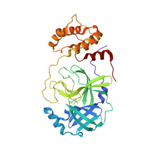
6XB1

Room temperature X-ray crystallography reveals catalytic cysteine in the SARS-CoV-2 3CL Mpro is highly reactive: Insights for enzyme mechanism and drug design


Experimental Data Snapshot

  • Method: X-RAY DIFFRACTION
  • Resolution: 1.80 Å
  • R-Value Free: 0.202 
  • R-Value Work: 0.159 
  • R-Value Observed: 0.161 

Starting Model: experimental
View more details

wwPDB Validation   3D Report Full Report


This is version 1.3 of the entry. See complete history


Literature

Room-temperature X-ray crystallography reveals the oxidation and reactivity of cysteine residues in SARS-CoV-2 3CL M pro : insights into enzyme mechanism and drug design.

Kneller, D.W.Phillips, G.O'Neill, H.M.Tan, K.Joachimiak, A.Coates, L.Kovalevsky, A.

(2020) IUCrJ 7

  • DOI: https://doi.org/10.1107/S2052252520012634
  • Primary Citation of Related Structures:  
    6XB0, 6XB1, 6XB2, 6XHU

  • PubMed Abstract: 

    The emergence of the novel coronavirus SARS-CoV-2 has resulted in a worldwide pandemic not seen in generations. Creating treatments and vaccines to battle COVID-19, the disease caused by the virus, is of paramount importance in order to stop its spread and save lives. The viral main protease, 3CL M pro , is indispensable for the replication of SARS-CoV-2 and is therefore an important target for the design of specific protease inhibitors. Detailed knowledge of the structure and function of 3CL M pro is crucial to guide structure-aided and computational drug-design efforts. Here, the oxidation and reactivity of the cysteine residues of the protease are reported using room-temperature X-ray crystallography, revealing that the catalytic Cys145 can be trapped in the peroxysulfenic acid oxidation state at physiological pH, while the other surface cysteines remain reduced. Only Cys145 and Cys156 react with the alkylating agent N -ethylmaleimide. It is suggested that the zwitterionic Cys145-His45 catalytic dyad is the reactive species that initiates catalysis, rather than Cys145-to-His41 proton transfer via the general acid-base mechanism upon substrate binding. The structures also provide insight into the design of improved 3CL M pro inhibitors.


  • Organizational Affiliation

    Neutron Scattering Division, Oak Ridge National Laboratory, 1 Bethel Valley Road, Oak Ridge, TN 37831, USA.


Macromolecules
Find similar proteins by:  (by identity cutoff)  |  3D Structure
Entity ID: 1
MoleculeChains Sequence LengthOrganismDetailsImage
3C-like proteinase306Severe acute respiratory syndrome coronavirus 2Mutation(s): 0 
EC: 3.4.22.69
UniProt
Find proteins for P0DTD1 (Severe acute respiratory syndrome coronavirus 2)
Explore P0DTD1 
Go to UniProtKB:  P0DTD1
Entity Groups  
Sequence Clusters30% Identity50% Identity70% Identity90% Identity95% Identity100% Identity
UniProt GroupP0DTD1
Sequence Annotations
Expand
  • Reference Sequence
Small Molecules
Ligands 1 Unique
IDChains Name / Formula / InChI Key2D Diagram3D Interactions
NEN
Query on NEN

Download Ideal Coordinates CCD File 
B [auth A]1-ETHYL-PYRROLIDINE-2,5-DIONE
C6 H9 N O2
GHAZCVNUKKZTLG-UHFFFAOYSA-N
Modified Residues  1 Unique
IDChains TypeFormula2D DiagramParent
2CO
Query on 2CO
A
L-PEPTIDE LINKINGC3 H7 N O4 SCYS
Experimental Data & Validation

Experimental Data

  • Method: X-RAY DIFFRACTION
  • Resolution: 1.80 Å
  • R-Value Free: 0.202 
  • R-Value Work: 0.159 
  • R-Value Observed: 0.161 
  • Space Group: I 1 2 1
Unit Cell:
Length ( Å )Angle ( ˚ )
a = 44.83α = 90
b = 53.89β = 100.338
c = 113.588γ = 90
Software Package:
Software NamePurpose
PHENIXrefinement
CrysalisProdata reduction
Aimlessdata scaling
MOLREPphasing

Structure Validation

View Full Validation Report


Entry History 

Deposition Data

Revision History  (Full details and data files)

  • Version 1.0: 2020-06-17
    Type: Initial release
  • Version 1.1: 2020-11-11
    Changes: Database references
  • Version 1.2: 2023-10-18
    Changes: Data collection, Database references, Refinement description
  • Version 1.3: 2024-10-23
    Changes: Structure summary