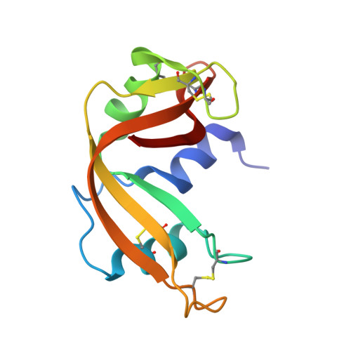
6XHD

Structure of Prolinyl-5'-O-adenosine phosphoramidate


Experimental Data Snapshot

  • Method: X-RAY DIFFRACTION
  • Resolution: 1.51 Å
  • R-Value Free: 0.217 
  • R-Value Work: 0.169 
  • R-Value Observed: 0.171 

Starting Model: experimental
View more details

wwPDB Validation   3D Report Full Report


Ligand Structure Quality Assessment 


This is version 1.1 of the entry. See complete history


Literature

The Enzyme-Free Release of Nucleotides from Phosphoramidates Depends Strongly on the Amino Acid.

Jovanovic, D.Tremmel, P.Pallan, P.S.Egli, M.Richert, C.

(2020) Angew Chem Int Ed Engl 59: 20154-20160

  • DOI: https://doi.org/10.1002/anie.202008665
  • Primary Citation of Related Structures:  
    6XHC, 6XHD, 6XHE, 6XHF

  • PubMed Abstract: 

    Phosphoramidates composed of an amino acid and a nucleotide analogue are critical metabolites of prodrugs, such as remdesivir. Hydrolysis of the phosphoramidate liberates the nucleotide, which can then be phosphorylated to become the pharmacologically active triphosphate. Enzymatic hydrolysis has been demonstrated, but a spontaneous chemical process may also occur. We measured the rate of enzyme-free hydrolysis for 17 phosphoramidates of ribonucleotides with amino acids or related compounds at pH 7.5. Phosphoramidates of proline hydrolyzed fast, with a half-life time as short as 2.4 h for Pro-AMP in ethylimidazole-containing buffer at 37 °C; 45-fold faster than Ala-AMP and 120-fold faster than Phe-AMP. Crystal structures of Gly-AMP, Pro-AMP, βPro-AMP and Phe-AMP bound to RNase A as crystallization chaperone showed how well the carboxylate is poised to attack the phosphoramidate, helping to explain this reactivity. Our results are significant for the design of new antiviral prodrugs.


  • Organizational Affiliation

    Institut für Organische Chemie, Universität Stuttgart, 70569, Stuttgart, Germany.


Macromolecules
Find similar proteins by:  (by identity cutoff)  |  3D Structure
Entity ID: 1
MoleculeChains Sequence LengthOrganismDetailsImage
Ribonuclease pancreatic
A, B
124Bos taurusMutation(s): 0 
Gene Names: RNASE1RNS1
EC: 4.6.1.18
UniProt
Find proteins for P61823 (Bos taurus)
Explore P61823 
Go to UniProtKB:  P61823
Entity Groups  
Sequence Clusters30% Identity50% Identity70% Identity90% Identity95% Identity100% Identity
UniProt GroupP61823
Sequence Annotations
Expand
  • Reference Sequence
Small Molecules
Ligands 3 Unique
IDChains Name / Formula / InChI Key2D Diagram3D Interactions
V2P (Subject of Investigation/LOI)
Query on V2P

Download Ideal Coordinates CCD File 
D [auth B](2~{S})-1-[[(2~{R},3~{S},4~{R},5~{R})-5-(6-aminopurin-9-yl)-3,4-bis(oxidanyl)oxolan-2-yl]methoxy-oxidanyl-phosphoryl]pyrrolidine-2-carboxylic acid
C15 H21 N6 O8 P
YHPWCYBNNLQQNG-TWBCTODHSA-N
MPD
Query on MPD

Download Ideal Coordinates CCD File 
E [auth B](4S)-2-METHYL-2,4-PENTANEDIOL
C6 H14 O2
SVTBMSDMJJWYQN-YFKPBYRVSA-N
K
Query on K

Download Ideal Coordinates CCD File 
C [auth A]POTASSIUM ION
K
NPYPAHLBTDXSSS-UHFFFAOYSA-N
Experimental Data & Validation

Experimental Data

  • Method: X-RAY DIFFRACTION
  • Resolution: 1.51 Å
  • R-Value Free: 0.217 
  • R-Value Work: 0.169 
  • R-Value Observed: 0.171 
  • Space Group: C 1 2 1
Unit Cell:
Length ( Å )Angle ( ˚ )
a = 98.532α = 90
b = 30.689β = 90.56
c = 68.641γ = 90
Software Package:
Software NamePurpose
REFMACrefinement
PDB_EXTRACTdata extraction
HKL-2000data reduction
HKL-2000data scaling
MOLREPphasing

Structure Validation

View Full Validation Report


Ligand Structure Quality Assessment 


Entry History & Funding Information

Deposition Data


Funding OrganizationLocationGrant Number
Volkswagen FoundationGermanygrant Az 92 768

Revision History  (Full details and data files)

  • Version 1.0: 2021-03-17
    Type: Initial release
  • Version 1.1: 2023-10-18
    Changes: Advisory, Data collection, Database references, Refinement description