7AA7

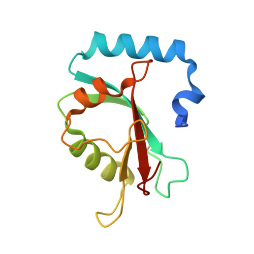
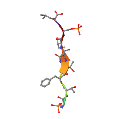
Structure of SCOC pS12/pS18 LIR motif bound to GABARAPL1


Experimental Data Snapshot

  • Method: X-RAY DIFFRACTION
  • Resolution: 1.45 Å
  • R-Value Free: 0.189 
  • R-Value Work: 0.151 
  • R-Value Observed: 0.153 

Starting Model: experimental
View more details

wwPDB Validation   3D Report Full Report


This is version 1.3 of the entry. See complete history


Literature

Phosphorylation of the LIR Domain of SCOC Modulates ATG8 Binding Affinity and Specificity.

Wirth, M.Mouilleron, S.Zhang, W.Sjottem, E.Princely Abudu, Y.Jain, A.Lauritz Olsvik, H.Bruun, J.A.Razi, M.Jefferies, H.B.J.Lee, R.Joshi, D.O'Reilly, N.Johansen, T.Tooze, S.A.

(2021) J Mol Biol 433: 166987-166987

  • DOI: https://doi.org/10.1016/j.jmb.2021.166987
  • Primary Citation of Related Structures:  
    7AA7, 7AA8, 7AA9

  • PubMed Abstract: 

    Autophagy is a highly conserved degradative pathway, essential for cellular homeostasis and implicated in diseases including cancer and neurodegeneration. Autophagy-related 8 (ATG8) proteins play a central role in autophagosome formation and selective delivery of cytoplasmic cargo to lysosomes by recruiting autophagy adaptors and receptors. The LC3-interacting region (LIR) docking site (LDS) of ATG8 proteins binds to LIR motifs present in autophagy adaptors and receptors. LIR-ATG8 interactions can be highly selective for specific mammalian ATG8 family members (LC3A-C, GABARAP, and GABARAPL1-2) and how this specificity is generated and regulated is incompletely understood. We have identified a LIR motif in the Golgi protein SCOC (short coiled-coil protein) exhibiting strong binding to GABARAP, GABARAPL1, LC3A and LC3C. The residues within and surrounding the core LIR motif of the SCOC LIR domain were phosphorylated by autophagy-related kinases (ULK1-3, TBK1) increasing specifically LC3 family binding. More distant flanking residues also contributed to ATG8 binding. Loss of these residues was compensated by phosphorylation of serine residues immediately adjacent to the core LIR motif, indicating that the interactions of the flanking LIR regions with the LDS are important and highly dynamic. Our comprehensive structural, biophysical and biochemical analyses support and provide novel mechanistic insights into how phosphorylation of LIR domain residues regulates the affinity and binding specificity of ATG8 proteins towards autophagy adaptors and receptors.


  • Organizational Affiliation

    Molecular Cell Biology of Autophagy, The Francis Crick Institute, 1 Midland Road, London NW1 1AT, UK; Department of Cell and Chemical Biology, Leiden University Medical Center, Einthovenweg 20, 2333 ZC Leiden, the Netherlands(‡). Electronic address: m.b.wirth@lumc.nl.


Macromolecules
Find similar proteins by:  (by identity cutoff)  |  3D Structure
Entity ID: 1
MoleculeChains Sequence LengthOrganismDetailsImage
Gamma-aminobutyric acid receptor-associated protein-like 1
A, B
123Homo sapiensMutation(s): 0 
Gene Names: GABARAPL1GEC1
UniProt & NIH Common Fund Data Resources
Find proteins for Q9H0R8 (Homo sapiens)
Explore Q9H0R8 
Go to UniProtKB:  Q9H0R8
PHAROS:  Q9H0R8
GTEx:  ENSG00000139112 
Entity Groups  
Sequence Clusters30% Identity50% Identity70% Identity90% Identity95% Identity100% Identity
UniProt GroupQ9H0R8
Sequence Annotations
Expand
  • Reference Sequence

Find similar proteins by:  Sequence   |   3D Structure  

Entity ID: 2
MoleculeChains Sequence LengthOrganismDetailsImage
pS12/pS18 SCOC LIRC [auth P],
D [auth Q]
11Homo sapiensMutation(s): 0 
UniProt & NIH Common Fund Data Resources
Find proteins for Q9UIL1 (Homo sapiens)
Explore Q9UIL1 
Go to UniProtKB:  Q9UIL1
PHAROS:  Q9UIL1
GTEx:  ENSG00000153130 
Entity Groups  
Sequence Clusters30% Identity50% Identity70% Identity90% Identity95% Identity100% Identity
UniProt GroupQ9UIL1
Sequence Annotations
Expand
  • Reference Sequence
Small Molecules
Modified Residues  1 Unique
IDChains TypeFormula2D DiagramParent
SEP
Query on SEP
C [auth P],
D [auth Q]
L-PEPTIDE LINKINGC3 H8 N O6 PSER
Experimental Data & Validation

Experimental Data

  • Method: X-RAY DIFFRACTION
  • Resolution: 1.45 Å
  • R-Value Free: 0.189 
  • R-Value Work: 0.151 
  • R-Value Observed: 0.153 
  • Space Group: P 1
Unit Cell:
Length ( Å )Angle ( ˚ )
a = 28.364α = 81.751
b = 37.169β = 89.602
c = 62.482γ = 67.818
Software Package:
Software NamePurpose
PHENIXrefinement
PHENIXrefinement
xia2data reduction
Aimlessdata scaling
PHASERphasing

Structure Validation

View Full Validation Report


Entry History 

Deposition Data

Revision History  (Full details and data files)

  • Version 1.0: 2021-06-16
    Type: Initial release
  • Version 1.1: 2023-01-18
    Changes: Database references
  • Version 1.2: 2024-01-31
    Changes: Data collection, Refinement description
  • Version 1.3: 2024-11-20
    Changes: Structure summary