7CUO | pdb_00007cuo

IclR transcription factor complexed with 4-hydroxybenzoic acid from Microbacterium hydrocarbonoxydans


Experimental Data Snapshot

  • Method: X-RAY DIFFRACTION
  • Resolution: 2.00 Å
  • R-Value Free: 
    0.218 (Depositor), 0.224 (DCC) 
  • R-Value Work: 
    0.202 (Depositor), 0.210 (DCC) 

Starting Model: experimental
View more details

wwPDB Validation 3D Report Full Report

Validation slider image for 7CUO

Ligand Structure Quality Assessment 


This is version 1.1 of the entry. See complete history

Literature

Structural basis of the conformational changes in Microbacterium hydrocarbonoxydans IclR transcription factor homolog due to ligand binding.

Akiyama, T.Sasaki, Y.Ito, S.Yajima, S.

(2021) Biochim Biophys Acta Proteins Proteom 1869: 140644-140644

  • DOI: https://doi.org/10.1016/j.bbapap.2021.140644
  • Primary Citation Related Structures: 
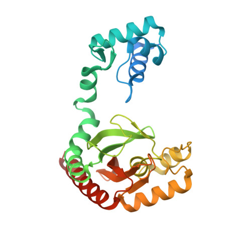
    7CUO, 7DQB

  • PubMed Abstract: 

    Microbacterium hydrocarbonoxydans has been isolated using an unnatural acylhydrazide compound as the sole carbon source. The compound is hydrolyzed by bacterial hydrazidase, and the gene expression of the enzyme is considered to be controlled by a transcription factor of the Isocitrate lyase Regulator (IclR) family, belonging to the one-component signaling systems. Recently, we reported the crystal structure of an unliganded IclR homolog from M. hydrocarbonoxydans, named putative 4-hydroxybenzoate response regulator (pHbrR), which has a unique homotetramer conformation. In this study, we report the crystal structure of pHbrR complexed with 4-hydroxybenzoic acid, the catalytic product of hydrazidase, at 2.0 Å resolution. pHbrR forms a homodimer with multimeric rearrangement in the unliganded state. Gel filtration column chromatography results suggested dimer-tetramer rearrangement. We observed conformational change in the loop region covering the ligand-binding site, and domain rearrangements in the monomer. This study reports the first liganded IclR family protein structure that demonstrates large structural rearrangements between liganded and unliganded proteins, which may represent a general model for IclRs.


  • Organizational Affiliation
    • Department of Bioscience, Tokyo University of Agriculture, Setagaya-ku, Tokyo 156-8502, Japan.

Macromolecule Content 

  • Total Structure Weight: 59.69 kDa 
  • Atom Count: 3,949 
  • Modeled Residue Count: 480 
  • Deposited Residue Count: 540 
  • Unique protein chains: 1

Macromolecules

Find similar proteins by:|  3D Structure
Entity ID: 1
MoleculeChains  Sequence LengthOrganismDetailsImage
Transcription factor
A, B
270Microbacterium hydrocarbonoxydansMutation(s): 0 

Small Molecules

Ligands 2 Unique
IDChains Name / Formula / InChI Key2D Diagram3D Interactions
PHB
(Subject of Investigation/LOI)

Query on PHB


Download:Ideal Coordinates CCD File
F [auth A],
L [auth B]
P-HYDROXYBENZOIC ACID
C7 H6 O3
FJKROLUGYXJWQN-UHFFFAOYSA-N
SO4

Query on SO4


Download:Ideal Coordinates CCD File
C [auth A]
D [auth A]
E [auth A]
G [auth B]
H [auth B]
C [auth A],
D [auth A],
E [auth A],
G [auth B],
H [auth B],
I [auth B],
J [auth B],
K [auth B]
SULFATE ION
O4 S
QAOWNCQODCNURD-UHFFFAOYSA-L

Experimental Data & Validation

Experimental Data

  • Method: X-RAY DIFFRACTION
  • Resolution: 2.00 Å
  • R-Value Free:  0.218 (Depositor), 0.224 (DCC) 
  • R-Value Work:  0.202 (Depositor), 0.210 (DCC) 
Space Group: P 41 21 2
Unit Cell:
Length ( Å )Angle ( ˚ )
a = 83.08α = 90
b = 83.08β = 90
c = 203.89γ = 90
Software Package:
Software NamePurpose
REFMACrefinement
XDSdata reduction
XSCALEdata scaling
MoRDaphasing

Structure Validation

View Full Validation Report


Ligand Structure Quality Assessment 


Entry History 

& Funding Information

Deposition Data


Funding OrganizationLocationGrant Number
Japan Society for the Promotion of Science (JSPS)Japan18K05398

Revision History  (Full details and data files)

  • Version 1.0: 2021-05-05
    Type: Initial release
  • Version 1.1: 2023-11-29
    Changes: Data collection, Database references, Derived calculations, Refinement description