7CZ0 | pdb_00007cz0

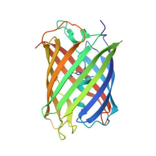
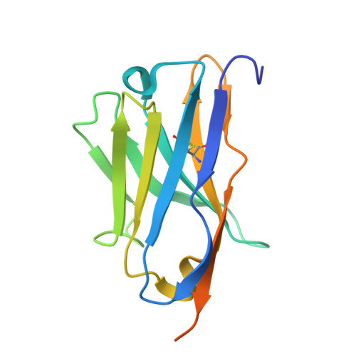
Crystal structure of a thermostable green fluorescent protein (TGP) with a synthetic nanobody (Sb92)


Experimental Data Snapshot

  • Method: X-RAY DIFFRACTION
  • Resolution: 2.77 Å
  • R-Value Free: 
    0.241 (Depositor), 0.241 (DCC) 
  • R-Value Work: 
    0.202 (Depositor), 0.204 (DCC) 
  • R-Value Observed: 
    0.204 (Depositor) 

Starting Model: experimental
View more details

wwPDB Validation 3D Report Full Report

Validation slider image for 7CZ0

This is version 3.1 of the entry. See complete history

Literature

Structure-based design of covalent nanobody binders for a thermostable green fluorescence protein.

Yue, Z.Li, Y.Cai, H.Yao, H.Li, D.Ni, A.Li, T.

(2024) Acta Biochim Biophys Sin (Shanghai) 57: 1363-1370

  • DOI: https://doi.org/10.3724/abbs.2024233
  • Primary Citation Related Structures: 
    7CZ0

  • PubMed Abstract: 

    The use of green fluorescence protein (GFP) has advanced numerous areas of life sciences. An ultra-thermostable GFP (TGP), engineered from a coral GFP, offers potential advantages over traditional jellyfish-derived GFP because of its high stability. However, owing to its later discovery, TGP lacks the extensive toolsets available for GFP, such as heavy chain-only antibody binders known as nanobodies. In this study, we report the crystal structure of TGP in complex with Sb92, a synthetic nanobody identified from a previous in vitro screening, revealing Sb92's precise three-dimensional epitope. This structural insight, alongside the previously characterized Sb44-TGP complex, allows us to rationally design disulfide bonds between the antigen and the antibody for tighter interactions. Using biochemical analysis, we identify two bridged complexes (TGP A18C-Sb44 V100C and TGP E118C-Sb92 S57C), with the TGP-Sb92 disulfide pair showing high resistance to reducing agents. Our study expands the toolkit available for TGP and should encourage its wider applications.


  • Organizational Affiliation
    • Center for Excellence in Molecular Cell Science, Shanghai Institute of Biochemistry and Cell Biology, Chinese Academy of Sciences, Shanghai 200031, China.

Macromolecule Content 

  • Total Structure Weight: 171.45 kDa 
  • Atom Count: 10,620 
  • Modeled Residue Count: 1,324 
  • Deposited Residue Count: 1,504 
  • Unique protein chains: 2

Macromolecules

Find similar proteins by:|  3D Structure
Entity ID: 1
MoleculeChains  Sequence LengthOrganismDetailsImage
Thermostable green fluorescent protein (TGP)
A, B, C, D
232Galaxea fascicularisMutation(s): 0 
Find similar proteins by:|  3D Structure
Entity ID: 2
MoleculeChains  Sequence LengthOrganismDetailsImage
Synthetic nanobody (Sybody) 92 recognizing the thermostable green fluorescent protein (TGP)
E, F, G, H
144synthetic constructMutation(s): 0 

Small Molecules

Ligands 4 Unique
IDChains Name / Formula / InChI Key2D Diagram3D Interactions
CAD

Query on CAD


Download:Ideal Coordinates CCD File
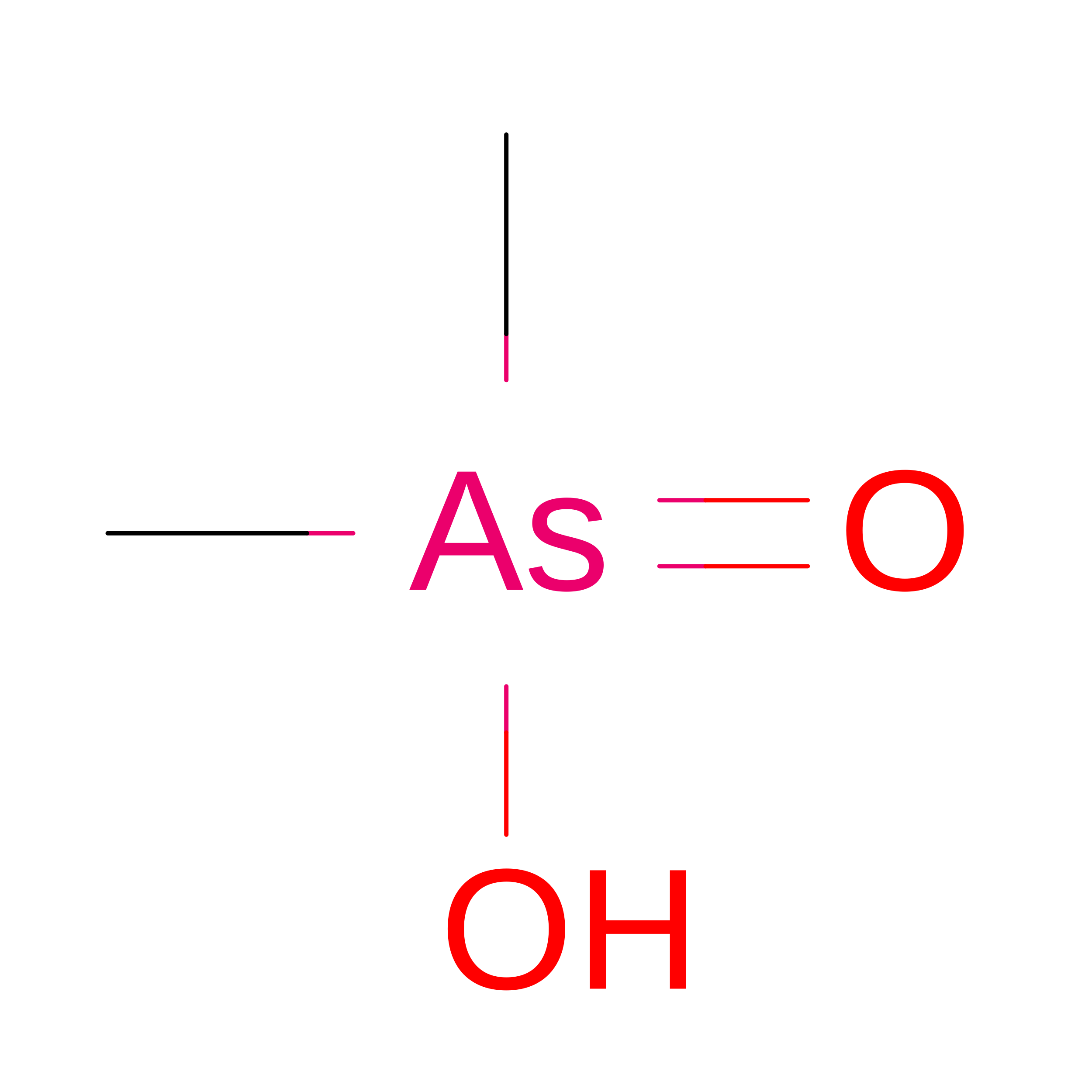
L [auth A]CACODYLIC ACID
C2 H7 As O2
OGGXGZAMXPVRFZ-UHFFFAOYSA-N
CAC

Query on CAC


Download:Ideal Coordinates CCD File
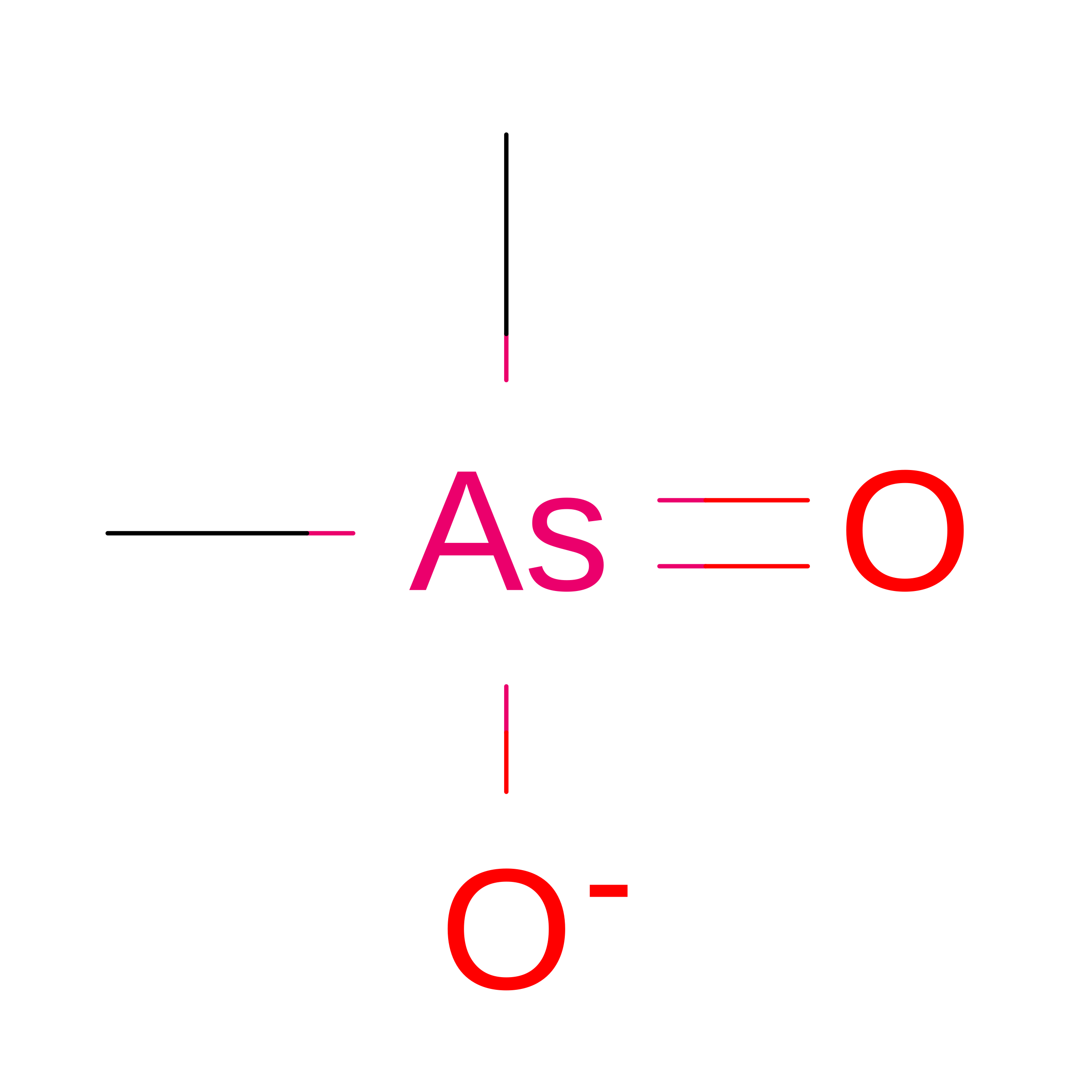
U [auth D]CACODYLATE ION
C2 H6 As O2
OGGXGZAMXPVRFZ-UHFFFAOYSA-M
GOL

Query on GOL


Download:Ideal Coordinates CCD File
I [auth A]
J [auth A]
K [auth A]
M [auth A]
N [auth B]
I [auth A],
J [auth A],
K [auth A],
M [auth A],
N [auth B],
O [auth B],
Q [auth B],
R [auth B],
S [auth B],
T [auth C],
V [auth D]
GLYCEROL
C3 H8 O3
PEDCQBHIVMGVHV-UHFFFAOYSA-N
ACT

Query on ACT


Download:Ideal Coordinates CCD File
P [auth B],
W [auth D]
ACETATE ION
C2 H3 O2
QTBSBXVTEAMEQO-UHFFFAOYSA-M
Modified Residues  1 Unique
IDChains TypeFormula2D DiagramParent
CRQ
Query on CRQ
A, B, C, D
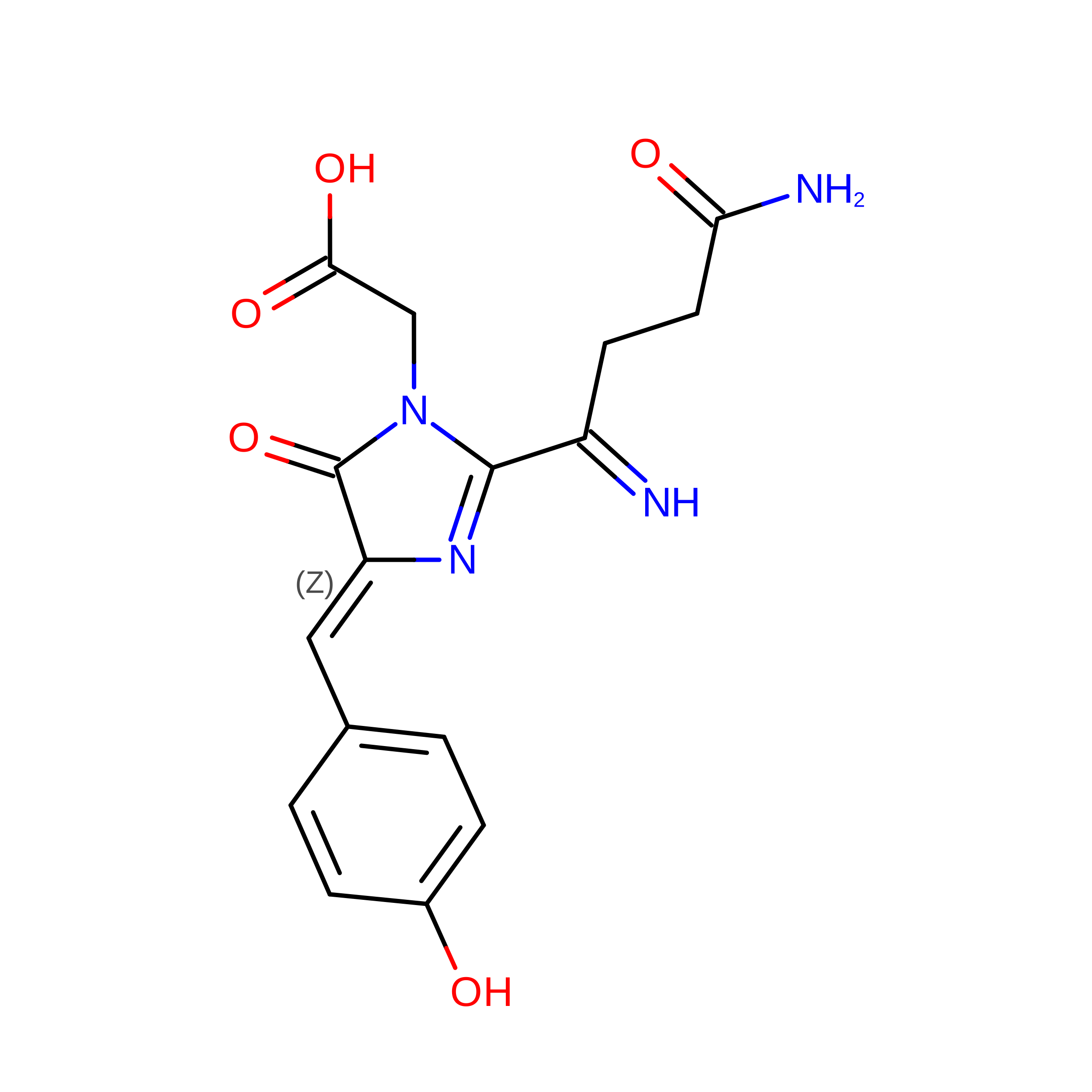
L-PEPTIDE LINKINGC16 H16 N4 O5GLN, TYR, GLY

Experimental Data & Validation

Experimental Data

  • Method: X-RAY DIFFRACTION
  • Resolution: 2.77 Å
  • R-Value Free:  0.241 (Depositor), 0.241 (DCC) 
  • R-Value Work:  0.202 (Depositor), 0.204 (DCC) 
  • R-Value Observed: 0.204 (Depositor) 
Space Group: P 21 21 2
Unit Cell:
Length ( Å )Angle ( ˚ )
a = 95.245α = 90
b = 130.154β = 90
c = 171.453γ = 90
Software Package:
Software NamePurpose
PHENIXrefinement
XDSdata reduction
Aimlessdata scaling
PHASERphasing

Structure Validation

View Full Validation Report


Entry History 

& Funding Information

Deposition Data


Funding OrganizationLocationGrant Number
National Science Foundation (NSF, China)China31870726

Revision History  (Full details and data files)

  • Version 1.0: 2021-09-08
    Type: Initial release
  • Version 2.0: 2023-11-15
    Changes: Atomic model, Data collection, Derived calculations
  • Version 2.1: 2023-11-29
    Changes: Refinement description
  • Version 2.2: 2024-10-16
    Changes: Structure summary
  • Version 3.0: 2026-03-18
    Changes: Polymer sequence
  • Version 3.1: 2026-07-01
    Changes: Database references