7DO6 | pdb_00007do6

Crystal structure of Azotobacter vinelandii L-rhamnose 1-dehydrogenase(NADP bound-form)


Experimental Data Snapshot

  • Method: X-RAY DIFFRACTION
  • Resolution: 2.37 Å
  • R-Value Free: 
    0.257 (Depositor), 0.255 (DCC) 
  • R-Value Work: 
    0.205 (Depositor), 0.203 (DCC) 
  • R-Value Observed: 
    0.208 (Depositor) 

wwPDB Validation 3D Report Full Report

Validation slider image for 7DO6

Ligand Structure Quality Assessment 


This is version 1.3 of the entry. See complete history

Literature

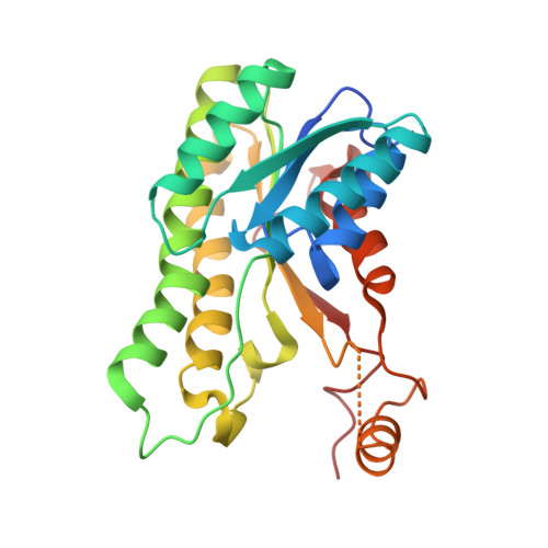
Crystal structure of l-rhamnose 1-dehydrogenase involved in the nonphosphorylative pathway of l-rhamnose metabolism in bacteria.

Yoshiwara, K.Watanabe, S.Watanabe, Y.

(2021) FEBS Lett 595: 637-646

  • DOI: https://doi.org/10.1002/1873-3468.14046
  • Primary Citation Related Structures: 
    7B81, 7DO5, 7DO6, 7DO7

  • PubMed Abstract: 

    Several microorganisms can utilize l-rhamnose as a carbon and energy source through the nonphosphorylative metabolic pathway, in which l-rhamnose 1-dehydrogenase (RhaDH) catalyzes the NAD(P) + -dependent oxidization of l-rhamnose to l-rhamnono-1,4-lactone. We herein investigated the crystal structures of RhaDH from Azotobacter vinelandii in ligand-free, NAD + -bound, NADP + -bound, and l-rhamnose- and NAD + -bound forms at 1.9, 2.1, 2.4, and 1.6 Å resolution, respectively. The significant interactions with the 2'-phosphate group of NADP + , but not the 2'-hydroxyl group of NAD + , were consistent with a preference for NADP + over NAD + . The C5-OH and C6-methyl groups of l-rhamnose were recognized by specific residues of RhaDH through hydrogen bonds and hydrophobic contact, respectively, which contribute to the different substrate specificities from other aldose 1-dehydrogenases in the short-chain dehydrogenase/reductase superfamily.


  • Organizational Affiliation
    • Faculty of Agriculture, Ehime University, Matsuyama, Japan.

Macromolecule Content 

  • Total Structure Weight: 227.71 kDa 
  • Atom Count: 14,756 
  • Modeled Residue Count: 1,994 
  • Deposited Residue Count: 2,136 
  • Unique protein chains: 1

Macromolecules

Find similar proteins by:|  3D Structure
Entity ID: 1
MoleculeChains  Sequence LengthOrganismDetailsImage
Short-chain dehydrogenase/reductase SDR
A, B, C, D, E
A, B, C, D, E, F, G, H
267Azotobacter vinelandii DJMutation(s): 0 
Gene Names: Avin_09160
EC: 1.1.1.378
UniProt
Find proteins for C1DMX5 (Azotobacter vinelandii (strain DJ / ATCC BAA-1303))
Explore C1DMX5 
Go to UniProtKB:  C1DMX5
Entity Groups
Sequence Clusters30% Identity50% Identity70% Identity90% Identity95% Identity100% Identity
UniProt GroupC1DMX5
Sequence Annotations
Expand
Reference Sequence

Small Molecules

Ligands 1 Unique
IDChains Name / Formula / InChI Key2D Diagram3D Interactions
NAP
(Subject of Investigation/LOI)

Query on NAP


Download:Ideal Coordinates CCD File
I [auth A]
J [auth B]
K [auth C]
L [auth D]
M [auth E]
I [auth A],
J [auth B],
K [auth C],
L [auth D],
M [auth E],
N [auth F],
O [auth G],
P [auth H]
NADP NICOTINAMIDE-ADENINE-DINUCLEOTIDE PHOSPHATE
C21 H28 N7 O17 P3
XJLXINKUBYWONI-NNYOXOHSSA-N

Experimental Data & Validation

Experimental Data

  • Method: X-RAY DIFFRACTION
  • Resolution: 2.37 Å
  • R-Value Free:  0.257 (Depositor), 0.255 (DCC) 
  • R-Value Work:  0.205 (Depositor), 0.203 (DCC) 
  • R-Value Observed: 0.208 (Depositor) 
Space Group: P 1
Unit Cell:
Length ( Å )Angle ( ˚ )
a = 78.633α = 87.68
b = 81.571β = 75.53
c = 86.389γ = 89.35
Software Package:
Software NamePurpose
PHENIXrefinement
XSCALEdata scaling
PDB_EXTRACTdata extraction
PHASERphasing

Structure Validation

View Full Validation Report


Ligand Structure Quality Assessment 


Entry History 

Deposition Data

Revision History  (Full details and data files)

  • Version 1.0: 2021-02-03
    Type: Initial release
  • Version 1.1: 2021-02-17
    Changes: Database references
  • Version 1.2: 2021-03-24
    Changes: Database references
  • Version 1.3: 2024-03-27
    Changes: Data collection, Database references