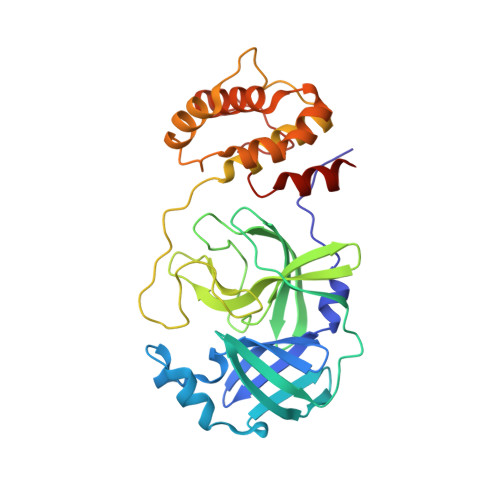
RCSB PDB - 7GRX: Crystal structure of SARS-CoV-2 main protease in complex with cpd-20

 7GRX

Crystal structure of SARS-CoV-2 main protease in complex with cpd-20


Experimental Data Snapshot

  • Method: X-RAY DIFFRACTION
  • Resolution: 1.55 Å
  • R-Value Free: 0.219 
  • R-Value Work: 0.187 
  • R-Value Observed: 0.188 

Starting Model: experimental
View more details

wwPDB Validation   3D Report Full Report


Ligand Structure Quality Assessment 

Created with Raphaël 2.3.0Worse 01 BetterLigand structure goodness of fit to experimental dataBest fitted XZTClick on this verticalbar to view details

This is version 1.1 of the entry. See complete history


Literature

Fragment-based screening targeting an open form of the SARS-CoV-2 main protease binding pocket.

Huang, C.Y.Metz, A.Lange, R.Artico, N.Potot, C.Hazemann, J.Muller, M.Dos Santos, M.Chambovey, A.Ritz, D.Eris, D.Meyer, S.Bourquin, G.Sharpe, M.Mac Sweeney, A.

(2024) Acta Crystallogr D Struct Biol 80: 123-136

  • DOI: https://doi.org/10.1107/S2059798324000329
  • Primary Citation of Related Structures:  
    7GRE, 7GRF, 7GRG, 7GRH, 7GRI, 7GRJ, 7GRK, 7GRL, 7GRM, 7GRN, 7GRO, 7GRP, 7GRQ, 7GRR, 7GRS, 7GRT, 7GRU, 7GRV, 7GRW, 7GRX, 7GRY, 7GRZ, 7GS0, 7GS1, 7GS2, 7GS3, 7GS4, 7GS5, 7GS6

  • PubMed Abstract: 

    To identify starting points for therapeutics targeting SARS-CoV-2, the Paul Scherrer Institute and Idorsia decided to collaboratively perform an X-ray crystallographic fragment screen against its main protease. Fragment-based screening was carried out using crystals with a pronounced open conformation of the substrate-binding pocket. Of 631 soaked fragments, a total of 29 hits bound either in the active site (24 hits), a remote binding pocket (three hits) or at crystal-packing interfaces (two hits). Notably, two fragments with a pose that was sterically incompatible with a more occluded crystal form were identified. Two isatin-based electrophilic fragments bound covalently to the catalytic cysteine residue. The structures also revealed a surprisingly strong influence of the crystal form on the binding pose of three published fragments used as positive controls, with implications for fragment screening by crystallography.


  • Organizational Affiliation

    Swiss Light Source, Paul Scherrer Institute, 5232 Villigen PSI, Switzerland.


Macromolecules
Find similar proteins by:  (by identity cutoff)  |  3D Structure
Entity ID: 1
MoleculeChains Sequence LengthOrganismDetailsImage
3C-like proteinase nsp5
A, B
306Severe acute respiratory syndrome coronavirus 2Mutation(s): 0 
Gene Names: rep1a-1b
EC: 3.4.22.69
UniProt
Find proteins for P0DTD1 (Severe acute respiratory syndrome coronavirus 2)
Explore P0DTD1 
Go to UniProtKB:  P0DTD1
Entity Groups  
Sequence Clusters30% Identity50% Identity70% Identity90% Identity95% Identity100% Identity
UniProt GroupP0DTD1
Sequence Annotations
Expand
  • Reference Sequence
Small Molecules
Ligands 4 Unique
IDChains Name / Formula / InChI Key2D Diagram3D Interactions
XZT (Subject of Investigation/LOI)
Query on XZT

Download Ideal Coordinates CCD File 
N [auth B]1-(2,4-difluorophenyl)pyrrolidine-2,5-dione
C10 H7 F2 N O2
YZJWAYPUZVVMIA-UHFFFAOYSA-N
DMS
Query on DMS

Download Ideal Coordinates CCD File 
C [auth A]
D [auth A]
E [auth A]
F [auth A]
H [auth A]
DIMETHYL SULFOXIDE
C2 H6 O S
IAZDPXIOMUYVGZ-UHFFFAOYSA-N
CL
Query on CL

Download Ideal Coordinates CCD File 
K [auth A],
Q [auth B]
CHLORIDE ION
Cl
VEXZGXHMUGYJMC-UHFFFAOYSA-M
NA
Query on NA

Download Ideal Coordinates CCD File 
G [auth A],
P [auth B]
SODIUM ION
Na
FKNQFGJONOIPTF-UHFFFAOYSA-N
Experimental Data & Validation

Experimental Data

  • Method: X-RAY DIFFRACTION
  • Resolution: 1.55 Å
  • R-Value Free: 0.219 
  • R-Value Work: 0.187 
  • R-Value Observed: 0.188 
  • Space Group: P 21 21 21
Unit Cell:
Length ( Å )Angle ( ˚ )
a = 67.87α = 90
b = 99.57β = 90
c = 103.83γ = 90
Software Package:
Software NamePurpose
PHENIXrefinement
XSCALEdata scaling
XDSdata reduction
PHASERphasing

Structure Validation

View Full Validation Report


Ligand Structure Quality Assessment 

Created with Raphaël 2.3.0Worse 01 BetterLigand structure goodness of fit to experimental dataBest fitted XZTClick on this verticalbar to view details

Entry History & Funding Information

Deposition Data


Funding OrganizationLocationGrant Number
Swiss National Science FoundationSwitzerland4078P0_198290

Revision History  (Full details and data files)

  • Version 1.0: 2024-02-14
    Type: Initial release
  • Version 1.1: 2024-03-13
    Changes: Source and taxonomy