A small molecule compound with an indole moiety inhibits the main protease of SARS-CoV-2 and blocks virus replication.
Hattori, S.I., Higashi-Kuwata, N., Hayashi, H., Allu, S.R., Raghavaiah, J., Bulut, H., Das, D., Anson, B.J., Lendy, E.K., Takamatsu, Y., Takamune, N., Kishimoto, N., Murayama, K., Hasegawa, K., Li, M., Davis, D.A., Kodama, E.N., Yarchoan, R., Wlodawer, A., Misumi, S., Mesecar, A.D., Ghosh, A.K., Mitsuya, H.(2021) Nat Commun 12: 668-668
- PubMed: 33510133 Search on PubMedSearch on PubMed Central
- DOI: https://doi.org/10.1038/s41467-021-20900-6
- Primary Citation Related Structures:
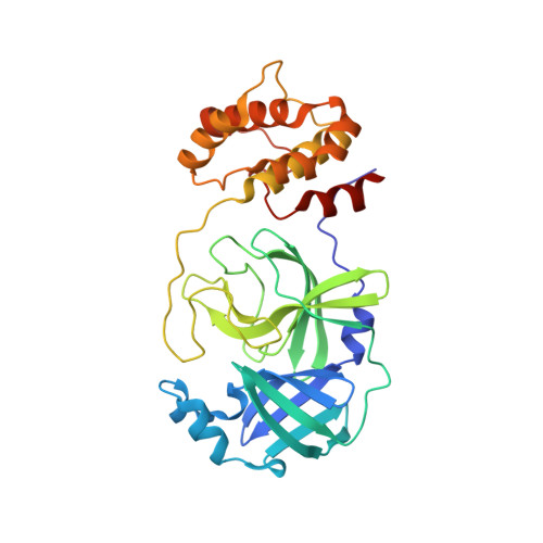
6XR3, 7JKV - PubMed Abstract:
Except remdesivir, no specific antivirals for SARS-CoV-2 infection are currently available. Here, we characterize two small-molecule-compounds, named GRL-1720 and 5h, containing an indoline and indole moiety, respectively, which target the SARS-CoV-2 main protease (M pro ). We use VeroE6 cell-based assays with RNA-qPCR, cytopathic assays, and immunocytochemistry and show both compounds to block the infectivity of SARS-CoV-2 with EC 50 values of 15 ± 4 and 4.2 ± 0.7 μM for GRL-1720 and 5h, respectively. Remdesivir permitted viral breakthrough at high concentrations; however, compound 5h completely blocks SARS-CoV-2 infection in vitro without viral breakthrough or detectable cytotoxicity. Combination of 5h and remdesivir exhibits synergism against SARS-CoV-2. Additional X-ray structural analysis show that 5h forms a covalent bond with M pro and makes polar interactions with multiple active site amino acid residues. The present data suggest that 5h might serve as a lead M pro inhibitor for the development of therapeutics for SARS-CoV-2 infection.
- Department of Refractory Viral Infections, National Center for Global Health and Medicine Research Institute, Tokyo, Japan.
Organizational Affiliation: