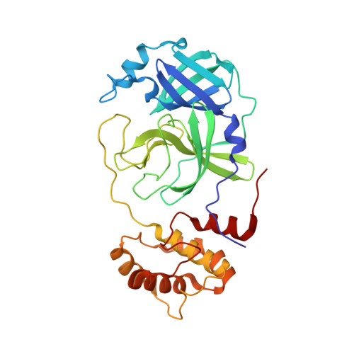
High-Throughput Virtual Screening and Validation of a SARS-CoV-2 Main Protease Noncovalent Inhibitor.
Clyde, A., Galanie, S., Kneller, D.W., Ma, H., Babuji, Y., Blaiszik, B., Brace, A., Brettin, T., Chard, K., Chard, R., Coates, L., Foster, I., Hauner, D., Kertesz, V., Kumar, N., Lee, H., Li, Z., Merzky, A., Schmidt, J.G., Tan, L., Titov, M., Trifan, A., Turilli, M., Van Dam, H., Chennubhotla, S.C., Jha, S., Kovalevsky, A., Ramanathan, A., Head, M.S., Stevens, R.(2022) J Chem Inf Model 62: 116-128
- PubMed: 34793155
- DOI: https://doi.org/10.1021/acs.jcim.1c00851
- Primary Citation of Related Structures:
7LTJ - PubMed Abstract:
Despite the recent availability of vaccines against the acute respiratory syndrome coronavirus 2 (SARS-CoV-2), the search for inhibitory therapeutic agents has assumed importance especially in the context of emerging new viral variants. In this paper, we describe the discovery of a novel noncovalent small-molecule inhibitor, MCULE-5948770040, that binds to and inhibits the SARS-Cov-2 main protease (M pro ) by employing a scalable high-throughput virtual screening (HTVS) framework and a targeted compound library of over 6.5 million molecules that could be readily ordered and purchased. Our HTVS framework leverages the U.S. supercomputing infrastructure achieving nearly 91% resource utilization and nearly 126 million docking calculations per hour. Downstream biochemical assays validate this M pro inhibitor with an inhibition constant ( K i ) of 2.9 μM (95% CI 2.2, 4.0). Furthermore, using room-temperature X-ray crystallography, we show that MCULE-5948770040 binds to a cleft in the primary binding site of M pro forming stable hydrogen bond and hydrophobic interactions. We then used multiple μs-time scale molecular dynamics (MD) simulations and machine learning (ML) techniques to elucidate how the bound ligand alters the conformational states accessed by M pro , involving motions both proximal and distal to the binding site. Together, our results demonstrate how MCULE-5948770040 inhibits M pro and offers a springboard for further therapeutic design.
- Data Science and Learning Division, Argonne National Laboratory, Lemont, Illinois 60439, United States.
Organizational Affiliation:


